

Mouse Monoclonal Antibody to CCRL2
Item Information
Catalog #
Size
Price
Description
This gene encodes a chemokine receptor like protein, which is predicted to be a seven transmembrane protein and most closely related to CCR1. Chemokines and their receptors mediated signal transduction are critical for the recruitment of effector immune cells to the site of inflammation. This gene is expressed at high levels in primary neutrophils and primary monocytes, and is further upregulated on neutrophil activation and during monocyte to macrophage differentiation. The function of this gene is unknown. This gene is mapped to the region where the chemokine receptor gene cluster is located.
Product Overview
Entrez GenelD
9034
Aliases
HCR;CKRX; CRAM;ACKR5; CRAM-A;
Clone#
4A5A5
Host / Isotype
Mouse / IgG1
Immunogen
Purified recombinant fragment of human CCRL2 expressed in E. Coli.
Formulation
Purified antibody in PBS with 0.05% sodium azide
Storage
4℃; -20℃ for long term storage
Product Applications
WB (Western Blot)
1/500 - 1/2000
FCM (Flow Cytometry)
1/200 - 1/400
ELISA
1/10000
References
1.Mol Immunol. 2015 Dec;68(2 Pt C):692-8. 2.Med Oncol. 2015 Nov;32(11):254.
Product Image
Elisa
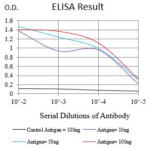
Figure 1:Black line: Control Antigen (100 ng);Purple line: Antigen (10ng); Blue line: Antigen (50 ng); Red line:Antigen (100 ng)
Western Blot
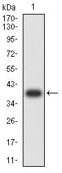
Figure 2:Western blot analysis using CCRL2 mAb against human CCRL2 recombinant protein. (Expected MW is 38.8 kDa)
Western Blot

Figure 3:Western blot analysis using CCRL2 mAb against HEK293-6e (1) and CCRL2-hIgGFc transfected HEK293-6e (2) cell lysate.
Flow cytometric analysis
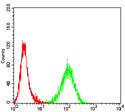
Figure 4:Flow cytometric analysis of Jurkat cells using CCRL2 mouse mAb (green) and negative control (red).
For Research Use Only. Not for use in diagnostic procedures.