
YWHAB Primary Antibody
Item Information
Catalog #
Size
Price
Description
This gene encodes a protein belonging to the 14-3-3 family of proteins, members of which mediate signal transduction by binding to phosphoserine-containing proteins. This highly conserved protein family is found in both plants and mammals. The encoded protein has been shown to interact with RAF1 and CDC25 phosphatases, suggesting that it may play a role in linking mitogenic signaling and the cell cycle machinery. Two transcript variants, which encode the same protein, have been identified for this gene.
Product Overview
Entrez GenelD
7529
Aliases
HS1; GW128; YWHAA; KCIP-1; HEL-S-1
Clone#
5B5G10
Host / Isotype
Mouse / IgG2b
Species Reactivity
Human, Mouse, Rat
Immunogen
Purified recombinant fragment of human YWHAB (AA: 1-246) expressed in E. Coli.
Formulation
Purified antibody in PBS with 0.05% sodium azide
Storage
Store at 4°C short term. Aliquot and store at -20°C long term. Avoid freeze/thaw cycles.
Product Applications
WB (Western Blot)
1/500 - 1/2000
IHC_P(Immunohistochemistry)
1/200 - 1/1000
ICC (Immunocytochemistry)
1/200 - 1/1000
FCM (Flow Cytometry)
1/200 - 1/400
ELISA
1/10000
References
1.BMC Res Notes. 2014 Feb 20;7:97.
2.Mol Biol Rep. 2012 Dec;39(12):10647-53.
2.Mol Biol Rep. 2012 Dec;39(12):10647-53.
Product Image
Western Blot
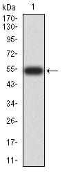
Figure 2:Western blot analysis using YWHAB mAb against human YWHAB (AA: 1-246) recombinant protein. (Expected MW is 54 kDa)
Western Blot
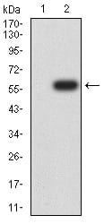
Figure 3:Western blot analysis using YWHAB mAb against HEK293 (1) and YWHAB (AA: 1-246)-hIgGFc transfected HEK293 (2) cell lysate.
Western Blot
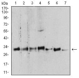
Figure 4:Western blot analysis using YWHAB mouse mAb against Hela (1), NIH/3T3 (2), C6 (3), A431 (4), K562 (5), PC-12 (6), and U937 (7) cell lysate.
Immunofluorescence analysis
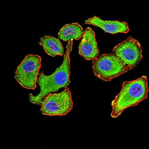
Figure 5:Immunofluorescence analysis of GC-7901 cells using YWHAB mouse mAb (green). Blue: DRAQ5 fluorescent DNA dye. Red: Actin filaments have been labeled with Alexa Fluor- 555 phalloidin. Secondary antibody from Fisher (Cat#: 35503)
Immunofluorescence analysis
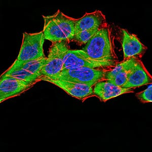
Figure 6:Immunofluorescence analysis of Hela cells using YWHAB mouse mAb (green). Blue: DRAQ5 fluorescent DNA dye. Red: Actin filaments have been labeled with Alexa Fluor- 555 phalloidin. Secondary antibody from Fisher (Cat#: 35503)
Immunofluorescence analysis
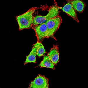
Figure 7:Immunofluorescence analysis of HepG2 cells using YWHAB mouse mAb (green). Blue: DRAQ5 fluorescent DNA dye. Red: Actin filaments have been labeled with Alexa Fluor- 555 phalloidin. Secondary antibody from Fisher (Cat#: 35503)
Flow cytometric
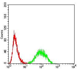
Figure 8:Flow cytometric analysis of Hela cells using YWHAB mouse mAb (green) and negative control (red).
Immunohistochemical analysis
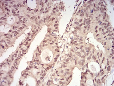
Figure 9:Immunohistochemical analysis of paraffin-embedded rectum cancer tissues using YWHAB mouse mAb with DAB staining.
Immunohistochemical analysis
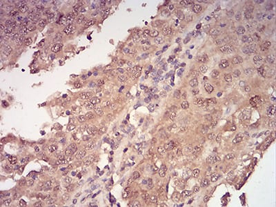
Figure 10:Immunohistochemical analysis of paraffin-embedded endometrial cancer tissues using YWHAB mouse mAb with DAB staining.
Elisa
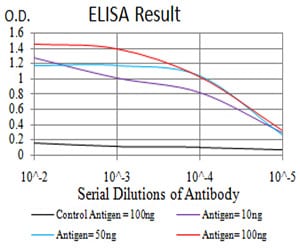
Black line: Control Antigen (100 ng);Purple line: Antigen (10ng); Blue line: Antigen (50 ng); Red line:Antigen (100 ng)
For Research Use Only. Not for use in diagnostic procedures.