
XRCC5 Primary Antibody
Item Information
Catalog #
Size
Price
Description
The protein encoded by this gene is the 80-kilodalton subunit of the Ku heterodimer protein which is also known as ATP-dependant DNA helicase II or DNA repair protein XRCC5. Ku is the DNA-binding component of the DNA-dependent protein kinase, and it functions together with the DNA ligase IV-XRCC4 complex in the repair of DNA double-strand break by non-homologous end joining and the completion of V(D)J recombination events. This gene functionally complements Chinese hamster xrs-6, a mutant defective in DNA double-strand break repair and in ability to undergo V(D)J recombination. A rare microsatellite polymorphism in this gene is associated with cancer in patients of varying radiosensitivity.
Product Overview
Entrez GenelD
7520
Aliases
KU80; KUB2; Ku86; NFIV; KARP1; KARP-1; FLJ39089
Clone#
5C5
Host / Isotype
Mouse / IgG1
Species Reactivity
Human, Mouse
Immunogen
Purified recombinant fragment of human XRCC5 expressed in E. Coli.
Formulation
Ascitic fluid containing 0.03% sodium azide.
Storage
Store at 4°C short term. Aliquot and store at -20°C long term. Avoid freeze/thaw cycles.
Product Applications
WB (Western Blot)
1/500 - 1/2000
IHC_P(Immunohistochemistry)
1/200 - 1/1000
ICC (Immunocytochemistry)
1/200 - 1/1000
FCM (Flow Cytometry)
1/200 - 1/400
ELISA
1/10000
References
1. Breast Cancer Res. 2009;11(6):R83.
2. Biochem Biophys Res Commun. 2009 Dec 18;390(3):738-42.
2. Biochem Biophys Res Commun. 2009 Dec 18;390(3):738-42.
Product Image
Western Blot
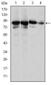
Figure 1: Western blot analysis using XRCC5 mouse mAb against Hela (1), MCF-7 (2), A549 (3) and NIH/3T3 (4) cell lysate.
Immunohistochemical analysis

Figure 2: Immunohistochemical analysis of paraffin-embedded human tonsil tissues (left) and human colon cancer tissues (right) using XRCC5 mouse mAb with DAB staining.
Immunofluorescence analysis

Figure 3: Immunofluorescence analysis of Hela cells using XRCC5 mouse mAb (green). Red: Actin filaments have been labeled with Alexa Fluor-555 phalloidin.
Flow cytometric

Figure 4: Flow cytometric analysis of Hela cells using XRCC5 mouse mAb (green) and negative control (purple).
Elisa

Red: Control Antigen (100ng); Purple: Antigen (10ng); Green: Antigen (50ng); Blue: Antigen (100ng);
For Research Use Only. Not for use in diagnostic procedures.