
VP2 Primary Antibody
Item Information
Catalog #
Size
Price
Description
Human parvovirus B19 (B19) is an erythrovirus responsible for acute and chronic anemia in susceptible patients. The virus replicates, both in vivo and in vitro, in the nucleus of the erythroid progenitors. The viral capsid is composed of two viral proteins, VP1 and VP2, the latter making up almost 96% of the viral capsid proteins
Product Overview
Entrez GenelD
11293627
Aliases
N
Clone#
4E5A4
Host / Isotype
Mouse / IgG1
Species Reactivity
Human
Immunogen
Purified recombinant fragment of human VP2 (AA: 296-438) expressed in E. Coli.
Formulation
Purified antibody in PBS with 0.05% sodium azide
Storage
Store at 4°C short term. Aliquot and store at -20°C long term. Avoid freeze/thaw cycles.
Product Applications
WB (Western Blot)
1/500 - 1/2000
IHC_P(Immunohistochemistry)
1/200 - 1/1000
ICC (Immunocytochemistry)
1/200 - 1/1000
FCM (Flow Cytometry)
1/200 - 1/400
ELISA
1/10000
References
1.Volume 306, Issue 1, 1 February 2003, Pages 25-32
Product Image
Elisa
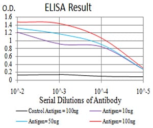
Figure 1: Black line: Control Antigen (100 ng);Purple line: Antigen (10ng); Blue line: Antigen (50 ng); Red line:Antigen (100 ng)
Western Blot
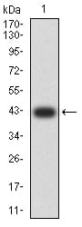
Figure 2:Western blot analysis using VP2 mAb against human VP2 (AA: 296-438) recombinant protein. (Expected MW is 42.1 kDa)
Western Blot
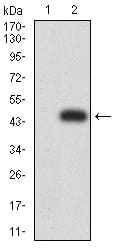
Figure 3:Western blot analysis using VP2 mAb against HEK293 (1) and VP2 (AA: 296-438)-hIgGFc transfected HEK293 (2) cell lysate.
Western Blot
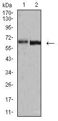
Figure 4:Western blot analysis using VP2 mouse mAb against A431 (1) and BCBL-1 (2) cell lysate.
Immunofluorescence analysis
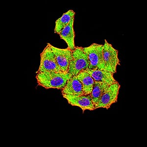
Figure 5:Immunofluorescence analysis of Hela cells using VP2 mouse mAb (green). Blue: DRAQ5 fluorescent DNA dye. Red: Actin filaments have been labeled with Alexa Fluor- 555 phalloidin. Secondary antibody from Fisher (Cat#: 35503)
Flow cytometric
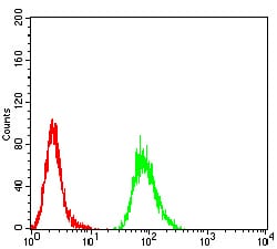
Figure 6:Flow cytometric analysis of Hela cells using VP2 mouse mAb (green) and negative control (red).
Immunohistochemical analysis
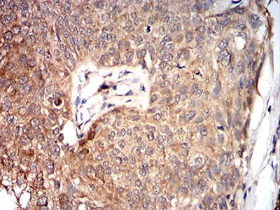
Figure 7:Immunohistochemical analysis of paraffin-embedded bladder cancer tissues using VP2 mouse mAb with DAB staining.
For Research Use Only. Not for use in diagnostic procedures.