
UHRF1 Primary Antibody
Item Information
Catalog #
Size
Price
Description
This gene encodes a member of a subfamily of RING-finger type E3 ubiquitin ligases. The protein binds to specific DNA sequences, and recruits a histone deacetylase to regulate gene expression. Its expression peaks at late G1 phase and continues during G2 and M phases of the cell cycle. It plays a major role in the G1/S transition by regulating topoisomerase IIalpha and retinoblastoma gene expression, and functions in the p53-dependent DNA damage checkpoint. It is regarded as a hub protein for the integration of epigenetic information. This gene is up-regulated in various cancers, and it is therefore considered to be a therapeutic target. Multiple transcript variants encoding different isoforms have been found for this gene. A related pseudogene exists on chromosome 12.
Product Overview
Entrez GenelD
29128
Aliases
Np95; hNP95; ICBP90; RNF106; TDRD22; hUHRF1; huNp95
Clone#
2A8C7
Host / Isotype
Mouse / IgG1
Species Reactivity
Human
Immunogen
Purified recombinant fragment of human UHRF1 (AA: 616-755) expressed in E. Coli.
Formulation
Purified antibody in PBS with 0.05% sodium azide
Storage
Store at 4°C short term. Aliquot and store at -20°C long term. Avoid freeze/thaw cycles.
Product Applications
WB (Western Blot)
1/500 - 1/2000
IHC_P(Immunohistochemistry)
1/200 - 1/1000
ELISA
1/10000
References
1.Biomarkers. 2015;20(3):183-8.
2.Med Oncol. 2013 Dec;30(4):613.
2.Med Oncol. 2013 Dec;30(4):613.
Product Image
Elisa
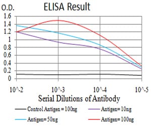
Figure 1: Black line: Control Antigen (100 ng);Purple line: Antigen (10ng); Blue line: Antigen (50 ng); Red line:Antigen (100 ng)
Western Blot
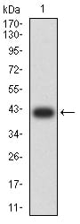
Figure 2:Western blot analysis using UHRF1 mAb against human UHRF1 (AA: 616-755) recombinant protein. (Expected MW is 41.8 kDa)
Western Blot
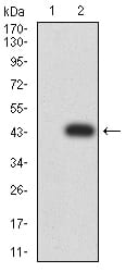
Figure 3:Western blot analysis using UHRF1 mAb against HEK293 (1) and UHRF1 (AA: 616-755)-hIgGFc transfected HEK293 (2) cell lysate.
Western Blot
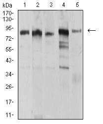
Figure 4:Western blot analysis using UHRF1 mouse mAb against MCF-7 (1), HCT116 (2), HL-60 (3), Hela (4), and HEK293 (5) cell lysate.
Immunohistochemical analysis
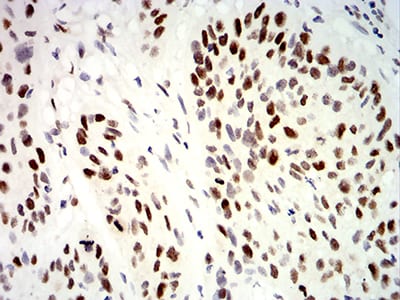
Figure 5:Immunohistochemical analysis of paraffin-embedded esophageal cancer tissues using UHRF1 mouse mAb with DAB staining.
For Research Use Only. Not for use in diagnostic procedures.