
TUBE1 Primary Antibody
Item Information
Catalog #
Size
Price
Description
This gene encodes a member of the tubulin superfamily. This protein localizes to the centriolar sub-distal appendages that are associated with the older of the two centrioles after centrosome duplication. This protein plays a central role in organization of the microtubules during centriole duplication. A pseudogene of this gene is found on chromosome 5.
Product Overview
Entrez GenelD
51175
Aliases
TUBE; dJ142L7.2
Clone#
5F3B7
Host / Isotype
Mouse / IgG1
Species Reactivity
Human
Immunogen
Purified recombinant fragment of human TUBE1 (AA: 314-472) expressed in E. Coli.
Formulation
Purified antibody from tissue culture in PBS with 0.05% sodium azide
Storage
Store at 4°C short term. Aliquot and store at -20°C long term. Avoid freeze/thaw cycles.
Product Applications
WB (Western Blot)
1/500 - 1/2000
IHC_P(Immunohistochemistry)
1/200 - 1/1000
FCM (Flow Cytometry)
1/200 - 1/400
ELISA
1/10000
References
1. Nat Cell Biol. 2003 Jan;5(1):71-6.
2. Nat Cell Biol. 2000 Jan;2(1):30-5.
2. Nat Cell Biol. 2000 Jan;2(1):30-5.
Product Image
Western Blot
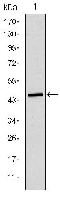
Figure 1: Western blot analysis using TUBE1 mAb against human TUBE1 (AA: 314-472) recombinant protein. (Expected MW is 44.3 kDa)
Western Blot
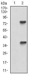
Figure 2: Western blot analysis using TUBE1 mAb against HEK293 (1) and TUBE1 (AA: 314-472)-hIgGFc transfected HEK293 (2) cell lysate.
Flow cytometric
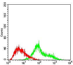
Figure 3: Flow cytometric analysis of Hela cells using TUBE1 mouse mAb (green) and negative control (red).
Immunohistochemical analysis
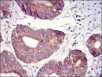
Figure 4: Immunohistochemical analysis of paraffin-embedded colon cancer tissues using TUBE1 mouse mAb with DAB staining.
Immunohistochemical analysis
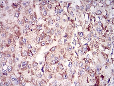
Figure 5: Immunohistochemical analysis of paraffin-embedded liver cancer tissues using TUBE1 mouse mAb with DAB staining.
Elisa
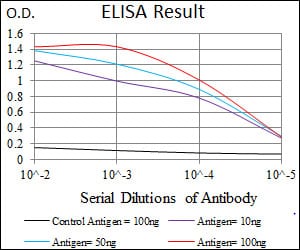
Black line: Control Antigen (100 ng); Purple line: Antigen(10ng); Blue line: Antigen (50 ng); Red line: Antigen (100 ng);
For Research Use Only. Not for use in diagnostic procedures.