
TSLPR Primary Antibody
Item Information
Catalog #
Size
Price
Description
This gene encodes a member of the type I cytokine receptor family. The encoded protein is a receptor for thymic stromal lymphopoietin (TSLP). Together with the interleukin 7 receptor (IL7R), the encoded protein and TSLP activate STAT3, STAT5, and JAK2 pathways, which control processes such as cell proliferation and development of the hematopoietic system. Rearrangement of this gene with immunoglobulin heavy chain gene (IGH) on chromosome 14, or with P2Y purinoceptor 8 gene (P2RY8) on the same X or Y chromosomes is associated with B-progenitor acute lymphoblastic leukemia (ALL) and Down syndrome ALL. Alternatively spliced transcript variants have been found for this gene.
Product Overview
Entrez GenelD
64109
Aliases
CRL2; TSLPR; CRLF2Y
Clone#
1E10D3
Host / Isotype
Mouse / Mouse IgG1
Immunogen
Purified recombinant fragment of human TSLPR (AA: extra(23-231)) expressed in E. Coli.
Formulation
Purified antibody in PBS with 0.05% sodium azide
Storage
Store at 4°C short term. Aliquot and store at -20°C long term. Avoid freeze/thaw cycles.
Product Applications
WB (Western Blot)
1/500 - 1/2000
IHC_P(Immunohistochemistry)
1/200-1/1000
FCM (Flow Cytometry)
1/200-1/400
ELISA
1/10000
References
1,PLoS One. 2019 Dec 12;14(12):e0224652.2,Zhongguo Shi Yan Xue Ye Xue Za Zhi. 2019 Aug;27(4):1058-1063.
Product Image
ELISA

Figure 1: Black line: Control Antigen (100 ng);Purple line: Antigen (10ng); Blue line: Antigen (50 ng); Red line: Antigen (100 ng)
WESTERN BLOT
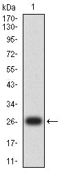
Figure 2: Western blot analysis using TSLPR mAb against human TSLPR (AA: extra(23-231)) recombinant protein. (Expected MW is 27 kDa)
WESTERN BLOT
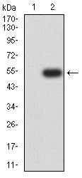
Figure 3: Western blot analysis using TSLPR mAb against HEK293-6e (1) and TSLPR (AA: extra(23-231))-hIgGFc transfected HEK293-6e (2) cell lysate.
FLOW CYTOMETRY
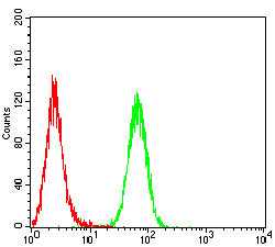
Figure 4: Flow cytometric analysis of THP-1 cells using TSLPR mouse mAb (green) and negative control (red).
IMMUNOHISTOCHEMISTRY
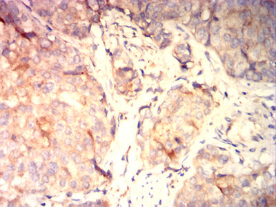
Figure 5: Immunohistochemical analysis of paraffin-embedded bladder cancer tissues using TSLPR mouse mAb with DAB staining.
IMMUNOHISTOCHEMISTRY
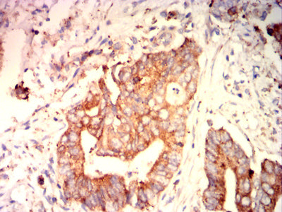
Figure 6: Immunohistochemical analysis of paraffin-embedded rectal cancer tissues using TSLPR mouse mAb with DAB staining.
For Research Use Only. Not for use in diagnostic procedures.