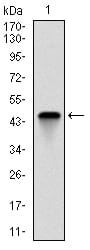
TNNI2 Primary Antibody

Item Information
Catalog #
Size
Price
Description

This gene encodes a fast-twitch skeletal muscle protein, a member of the troponin I gene family, and a component of the troponin complex including troponin T, troponin C and troponin I subunits. The troponin complex, along with tropomyosin, is responsible for the calcium-dependent regulation of striated muscle contraction. Mouse studies show that this component is also present in vascular smooth muscle and may play a role in regulation of smooth muscle function. In addition to muscle tissues, this protein is found in corneal epithelium, cartilage where it is an inhibitor of angiogenesis to inhibit tumor growth and metastasis, and mammary gland where it functions as a co-activator of estrogen receptor-related receptor alpha. This protein also suppresses tumor growth in human ovarian carcinoma. Mutations in this gene cause myopathy and distal arthrogryposis type 2B. Alternatively spliced transcript variants have been found for this gene.

Product Overview
Entrez GenelD
7136
Aliases
DA2B; FSSV; fsTnI; AMCD2B
Clone#
2F12G2
Host / Isotype
Mouse / IgG1
Species Reactivity
Human
Immunogen
Purified recombinant fragment of human TNNI2 expressed in E. Coli.
Formulation
Purified antibody in PBS with 0.05% sodium azide
Storage
4°C; -20°C for long term storage
Product Applications
WB (Western Blot)
1/500 - 1/2000
ICC (Immunocytochemistry)
1/200 - 1/1000
FCM (Flow Cytometry)
1/200 - 1/400
ELISA
1/10000
References
1. Am J Hum Genet. 2009 Nov;85(5):628-42.
2. Cell Motil Cytoskeleton. 2008 Aug;65(8):652-61.
Product Image
Western Blot
Figure 1: Western blot analysis using TNNI2 mAb against human TNNI2 (AA: 1-182) recombinant protein. (Expected MW is 21 kDa)
Western Blot
Figure 2: Western blot analysis using TNNI2 mAb against HEK293 (1) and TNNI2 (AA: 1-182)-hIgGFc transfected HEK293 (2) cell lysate.
Immunofluorescence analysis
Figure 3: Immunofluorescence analysis of Hela cells using TNNI2 mouse mAb (green). Blue: DRAQ5 fluorescent DNA dye. Red: Actin filaments have been labeled with Alexa Fluor-555 phalloidin.
Flow cytometric
Figure 4: Flow cytometric analysis of NIH/3T3 cells using TNNI2 mouse mAb (green) and negative control (red).
Elisa
Black line: Control Antigen (100 ng); Purple line: Antigen(10ng); Blue line: Antigen (50 ng); Red line: Antigen (100 ng);
For Research Use Only. Not for use in diagnostic procedures.