
THY1 Primary Antibody
Item Information
Catalog #
Size
Price
Description
THY1 may play a role in cell-cell or cell-ligand interactions during synaptogenesis and other events in the brain
Product Overview
Entrez GenelD
7070
Aliases
CD90
Clone#
7E1B11
Host / Isotype
Mouse / IgG1
Species Reactivity
Human
Immunogen
Purified recombinant fragment of human THY1 (AA: 17-132) expressed in E. Coli.
Formulation
Purified antibody in PBS with 0.05% sodium azide
Storage
Store at 4°C short term. Aliquot and store at -20°C long term. Avoid freeze/thaw cycles.
Product Applications
WB (Western Blot)
1/500 - 1/2000
IHC_P(Immunohistochemistry)
1/200 - 1/1000
ICC (Immunocytochemistry)
1/200 - 1/1000
ELISA
1/10000
References
1. Acta Histochem. 2011 Dec;113(8):833-8.
2. J Immunol. 2012 Feb 1;188(3):981-91.
2. J Immunol. 2012 Feb 1;188(3):981-91.
Product Image
Western Blot

Figure 1: Western blot analysis using THY1 mAb against human THY1 recombinant protein. (Expected MW is 38.5 kDa)
Western Blot

Figure 2: Western blot analysis using THY1 mAb against HEK293 (1) and THY1 (AA: 17-132)-hIgGFc transfected HEK293 (2) cell lysate.
Western Blot

Figure 3: Western blot analysis using THY1 mouse mAb against T47D (1), HepG2 (2) and PC-12 (3) cell lysate.
Immunofluorescence analysis

Figure 4: Immunofluorescence analysis of Hela cells using THY1 mouse mAb (green). Blue: DRAQ5 fluorescent DNA dye. Red: Actin filaments have been labeled with Alexa Fluor-555 phalloidin.
Immunohistochemical analysis

Figure 5: Immunohistochemical analysis of paraffin-embedded endometrial cancer tissues using THY1 mouse mAb with DAB staining.
Immunohistochemical analysis

Figure 6: Immunohistochemical analysis of paraffin-embedded cerebellum tissues using THY1 mouse mAb with DAB staining.
Elisa
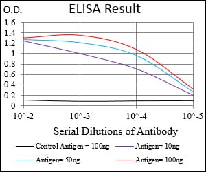
Black line: Control Antigen (100 ng); Purple line: Antigen(10ng); Blue line: Antigen (50 ng); Red line: Antigen (100 ng);
For Research Use Only. Not for use in diagnostic procedures.