
SYCP3 Primary Antibody
Item Information
Catalog #
Size
Price
Description
This gene encodes an essential structural component of the synaptonemal complex. This complex is involved in synapsis, recombination and segregation of meiotic chromosomes. Mutations in this gene are associated with azoospermia in males and susceptibility to pregnancy loss in females. Alternate splicing results in multiple transcript variants that encode the same protein.
Product Overview
Entrez GenelD
50511
Aliases
COR1; SCP3; SPGF4
Clone#
6F9C5
Host / Isotype
Mouse / IgG1
Species Reactivity
Human
Immunogen
Purified recombinant fragment of human SYCP3 (AA: 27-128) expressed in E. Coli.
Formulation
Purified antibody in PBS with 0.05% sodium azide
Storage
Store at 4°C short term. Aliquot and store at -20°C long term. Avoid freeze/thaw cycles.
Product Applications
WB (Western Blot)
1/500 - 1/2000
IHC_P(Immunohistochemistry)
1/200 - 1/1000
ICC (Immunocytochemistry)
1/200 - 1/1000
FCM (Flow Cytometry)
1/200 - 1/400
ELISA
1/10000
References
1. Hum Pathol. 2013 Apr;44(4):472-9.
2. Cytogenet Genome Res. 2010;128(1-3):162-8.
2. Cytogenet Genome Res. 2010;128(1-3):162-8.
Product Image
Western Blot

Figure 1: Western blot analysis using SYCP3 mAb against human SYCP3 recombinant protein. (Expected MW is 37.2 kDa)
Western Blot

Figure 2: Western blot analysis using SYCP3 mAb against HEK293 (1) and SYCP3 (AA: 27-128)-hIgGFc transfected HEK293 (2) cell lysate.
Immunofluorescence analysis

Figure 3: Immunofluorescence analysis of HepG2 cells using SYCP3 mouse mAb (green). Blue: DRAQ5 fluorescent DNA dye. Red: Actin filaments have been labeled with Alexa Fluor-555 phalloidin.
Flow cytometric

Figure 4: Flow cytometric analysis of Jurkat cells using SYCP3 mouse mAb (green) and negative control (red).
Immunohistochemical analysis

Figure 5: Immunohistochemical analysis of paraffin-embedded cervical cancer tissues using SYCP3 mouse mAb with DAB staining.
Immunohistochemical analysis
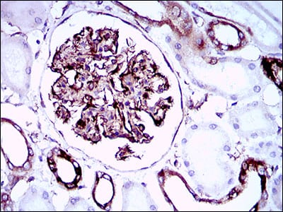
Figure 6: Immunohistochemical analysis of paraffin-embedded kidney tissues using SYCP3 mouse mAb with DAB staining.
Elisa

Black line: Control Antigen (100 ng); Purple line: Antigen(10ng); Blue line: Antigen (50 ng); Red line: Antigen (100 ng);
For Research Use Only. Not for use in diagnostic procedures.