
SMAD5 Primary Antibody
Item Information
Catalog #
Size
Price
Description
Transcriptional modulator activated by BMP (bone morphogenetic proteins) type 1 receptor kinase. SMAD5 is a receptor-regulated SMAD (R-SMAD). SMAD5 is required for normal development of the cardiovascular system in vivo; lack of the SMAD5 gene results in apoptosis of cardiac myocytes. 3 Upregulation of SMAD5 has been reported to mediate apoptosis of gastric epithelial cells induced by Helicobacter pylori infection. Tissue specificity: Ubiquitous.
Product Overview
Entrez GenelD
4090
Aliases
Dwfc; JV5-1; MADH5; DKFZp781C1895; DKFZp781O1323; SMAD5
Clone#
3H9
Host / Isotype
Mouse / IgG1
Species Reactivity
Human, Rat
Immunogen
Purified recombinant fragment of human SMAD5 expressed in E. Coli.
Formulation
Ascitic fluid containing 0.03% sodium azide.
Storage
Store at 4°C short term. Aliquot and store at -20°C long term. Avoid freeze/thaw cycles.
Product Applications
WB (Western Blot)
1/500 - 1/2000
IHC_P(Immunohistochemistry)
1/200 - 1/1000
ICC (Immunocytochemistry)
1/200 - 1/1000
FCM (Flow Cytometry)
1/200 - 1/400
ELISA
1/10000
References
1. Proc Natl Acad Sci U S A. 2008 Mar 11;105(10):3927-32.
2. Nat Cell Biol. 2008 May;10(5):567-74.
2. Nat Cell Biol. 2008 May;10(5):567-74.
Product Image
Western Blot
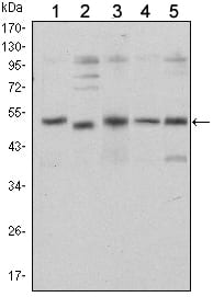
Figure 1: Western blot analysis using SMAD5 mouse mAb against Hela (1), SK-N-SH (2), PC-12 (3), Jurkat (4), and K562 (5) cell lysate.
Immunohistochemical analysis

Figure 2: Immunohistochemical analysis of paraffin-embedded brain tissues (left) and lung cancer tissues (right) using SMAD5 mouse mAb with DAB staining.
Immunofluorescence analysis

Figure 3: Immunofluorescence analysis of NTERA-2 cells using SMAD5 mouse mAb (green). Blue: DRAQ5 fluorescent DNA dye. Red: Actin filaments have been labeled with Alexa Fluor-555 phalloidin.
Flow cytometric

Figure 4: Flow cytometric analysis of Jurkat cells using SMAD5 mouse mAb (green) and negative control (purple).
For Research Use Only. Not for use in diagnostic procedures.