
Mouse Monoclonal Antibody to SIGLEC8
Item Information
Catalog #
Size
Price
Description
Sialic acid-binding immunoglobulin (Ig)-like lectins, or SIGLECs (e.g., CD33 (MIM 159590)), are a family of type 1 transmembrane proteins each having a unique expression pattern, mostly in hemopoietic cells. SIGLEC8 is a member of the CD33-like subgroup of SIGLECs, which are localized to 19q13.3-q13.4 and have 2 conserved cytoplasmic tyrosine-based motifs: an immunoreceptor tyrosine-based inhibitory motif, or ITIM (see MIM 604964), and a motif homologous to one identified in signaling lymphocyte activation molecule (SLAM; MIM 603492) that mediates an association with SLAM-associated protein (SAP; MIM 300490) (summarized by Foussias et al., 2000 [PubMed 11095983])
Product Overview
Entrez GenelD
27181
Aliases
SAF2; SIGLEC-8; SIGLEC8L
Clone#
1A5B9
Host / Isotype
Mouse / IgG2b
Immunogen
Purified recombinant fragment of human SIGLEC8 (AA: extra 17-216) expressed in E. Coli.
Formulation
Purified antibody in PBS with 0.05% sodium azide
Storage
Store at 4°C short term. Aliquot and store at -20°C long term. Avoid freeze/thaw cycles.
Product Applications
WB (Western Blot)
1/500 - 1/2000
IHC_P(Immunohistochemistry)
1/200 - 1/1000
FCM (Flow Cytometry)
1/200 - 1/400
ELISA
1/10000
References
1.J Allergy Clin Immunol. 2019 Jun;143(6):2227-2237.e10. 2.Tumour Biol. 2016 Aug;37(8):10883-91.
Product Image
Elisa
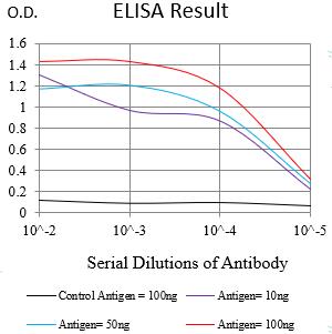
Figure 1:Black line: Control Antigen (100 ng);Purple line: Antigen (10ng); Blue line: Antigen (50 ng); Red line:Antigen (100 ng)
Western Blot
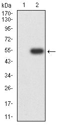
Figure 2:Western blot analysis using SIGLEC8 mAb against human SIGLEC8 (AA: extra 17-216) recombinant protein. (Expected MW is 25.3 kDa)
Western Blot

Figure 3:Western blot analysis using SIGLEC8 mAb against HEK293-6e (1) and SIGLEC8 (AA: extra 17-216)-hIgGFc transfected HEK293-6e (2) cell lysate.
Western Blot
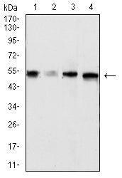
Figure 4:Western blot analysis using SIGLEC8 mouse mAb against mouse Liver (1), rat Liver (2)tissues lysate, MCF-7 (3), and HT-29 (4) cell lysate.
Flow cytometric analysis
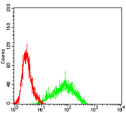
Figure 5:Flow cytometric analysis of Jurkat cells using SIGLEC8 mouse mAb (green) and negative control (red).
Immunohistochemical analysis
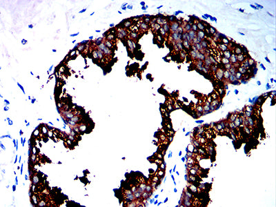
Figure 6:Immunohistochemical analysis of paraffin-embedded prostate cancer tissues using SIGLEC8 mouse mAb with DAB staining.
Immunohistochemical analysis
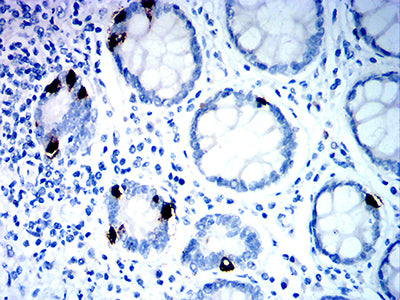
Figure 7:Immunohistochemical analysis of paraffin-embedded rectum tissues using SIGLEC8 mouse mAb with DAB staining.
For Research Use Only. Not for use in diagnostic procedures.