
SERPINA7 Primary Antibody
Item Information
Catalog #
Size
Price
Description
There are three proteins including thyroxine-binding globulin (TBG), transthyretin and albumin responsible for carrying the thyroid hormones thyroxine (T4) and 3,5,3'-triiodothyronine (T3) in the bloodstream. This gene encodes the major thyroid hormone transport protein, TBG, in serum. It belongs to the serpin family in genomics, but the protein has no inhibitory function like many other members of the serpin family. Mutations in this gene result in TGB deficiency, which has been classified as partial deficiency, complete deficiency, and excess, based on the level of serum TBG. Alternatively spliced transcript variants encoding different isoforms have been found, but the full-length nature of these variants has not been determined.
Product Overview
Entrez GenelD
6906
Aliases
TBG
Clone#
5B11E9
Host / Isotype
Mouse / IgG1
Species Reactivity
Human
Immunogen
Purified recombinant fragment of human SERPINA7 (AA: 168-302) expressed in E. Coli.
Formulation
Purified antibody in PBS with 0.05% sodium azide
Storage
Store at 4°C short term. Aliquot and store at -20°C long term. Avoid freeze/thaw cycles.
Product Applications
WB (Western Blot)
1/500 - 1/2000
IHC_P(Immunohistochemistry)
1/200 - 1/1000
ICC (Immunocytochemistry)
1/200 - 1/1000
FCM (Flow Cytometry)
1/200 - 1/400
ELISA
1/10000
References
1. Gene. 2012 Sep 15;506(2):289-94.
2. Endocr Regul. 2010 Apr;44(2):43-7.
2. Endocr Regul. 2010 Apr;44(2):43-7.
Product Image
Western Blot
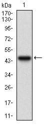
Figure 1: Western blot analysis using SERPINA7 mAb against human SERPINA7 recombinant protein. (Expected MW is 41.4 kDa)
Western Blot

Figure 2: Western blot analysis using SERPINA7 mAb against HEK293 (1) and SERPINA7 (AA: 168-302)-hIgGFc transfected HEK293 (2) cell lysate.
Immunofluorescence analysis

Figure 3: Immunofluorescence analysis of A431 cells using SERPINA7 mouse mAb (green). Blue: DRAQ5 fluorescent DNA dye.
Flow cytometric

Figure 4: Flow cytometric analysis of A431 cells using SERPINA7 mouse mAb (green) and negative control (red).
Immunohistochemical analysis

Figure 5: Immunohistochemical analysis of paraffin-embedded liver cancer tissues using SERPINA7 mouse mAb with DAB staining.
Immunohistochemical analysis

Figure 6: Immunohistochemical analysis of paraffin-embedded bladder cancer tissues using SERPINA7 mouse mAb with DAB staining.
Elisa

Black line: Control Antigen (100 ng); Purple line: Antigen(10ng); Blue line: Antigen (50 ng); Red line: Antigen (100 ng);
For Research Use Only. Not for use in diagnostic procedures.