
SDHA Primary Antibody
Item Information
Catalog #
Size
Price
Description
This gene encodes a major catalytic subunit of succinate-ubiquinone oxidoreductase, a complex of the mitochondrial respiratory chain. The complex is composed of four nuclear-encoded subunits and is localized in the mitochondrial inner membrane. Mutations in this gene have been associated with a form of mitochondrial respiratory chain deficiency known as Leigh Syndrome. A pseudogene has been identified on chromosome 3q29. Alternatively spliced transcript variants encoding different isoforms have been found for this gene.
Product Overview
Entrez GenelD
6389
Aliases
FP; PGL5; SDH1; SDH2; SDHF; CMD1GG; MC2DN1; NDAXOA
Clone#
2A10F6
Host / Isotype
Mouse / Mouse IgG2a
Species Reactivity
Human, Mouse
Immunogen
Purified recombinant fragment of human SDHA (AA: 516-665) expressed in E. Coli.
Formulation
Purified antibody in PBS with 0.05% sodium azide
Storage
Store at 4°C short term. Aliquot and store at -20°C long term. Avoid freeze/thaw cycles.
Product Applications
WB (Western Blot)
1/500 - 1/2000
ICC (Immunocytochemistry)
1/200 - 1/1000
FCM (Flow Cytometry)
1/200 - 1/400
ELISA
1/10000
References
1,Cancer Sci . 2021 Aug;112(8):3375-3387.
2,Medicine (Baltimore) . 2020 Oct 9;99(41):e22497.
2,Medicine (Baltimore) . 2020 Oct 9;99(41):e22497.
Product Image
Elisa

Figure 1:Black line: Control Antigen (100 ng);Purple line: Antigen (10ng); Blue line: Antigen (50 ng); Red line:Antigen (100 ng)
Western Blot
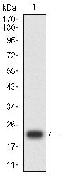
Figure 2:Western blot analysis using SDHA mAb against human SDHA (AA: 516-665) recombinant protein. (Expected MW is 20 kDa)
Western Blot

Figure 3:Western blot analysis using SDHA mAb against HEK293-6e (1) and SDHA (AA: 516-665)-hIgGFc transfected HEK293-6e (2) cell lysate.
Western Blot

Figure 4:Western blot analysis using SDHA mouse mAb against Jurkat (1), MCF-7 (2), Hela (3), HepG2 (4), PC-3 (5), .HL-60 (6),and NIH/3T3 (7) cell lysate.
Immunohistochemical analysis

Figure 5:Immunofluorescence analysis of Hela cells using SDHA mouse mAb (green). Blue: DRAQ5 fluorescent DNA dye. Red: Actin filaments have been labeled with Alexa Fluor- 555 phalloidin. Secondary antibody from Fisher (Cat#: 35503)
Immunofluorescence analysis

Figure 6:Flow cytometric analysis of Raji cells using SDHA mouse mAb (green) and negative control (red).
Immunofluorescence analysis

Figure 7:Flow cytometric analysis of THP-1 cells using SDHA mouse mAb (green) and negative control (red).
For Research Use Only. Not for use in diagnostic procedures.