
PRDM5 Primary Antibody
Item Information
Catalog #
Size
Price
Description
The protein encoded by this gene is a transcription factor of the PR-domain protein family. It contains a PR-domain and multiple zinc finger motifs. Transcription factors of the PR-domain family are known to be involved in cell differentiation and tumorigenesis.
Product Overview
Entrez GenelD
11107
Aliases
BCS2; PFM2
Clone#
7D4C12
Host / Isotype
Mouse / IgG1
Species Reactivity
Human, Mouse
Immunogen
Purified recombinant fragment of human PRDM5 (AA: 1-100) expressed in E. Coli.
Formulation
Purified antibody in PBS with 0.05% sodium azide
Storage
Store at 4°C short term. Aliquot and store at -20°C long term. Avoid freeze/thaw cycles.
Product Applications
WB (Western Blot)
1/500 - 1/2000
ICC (Immunocytochemistry)
1/100 - 1/500
FCM (Flow Cytometry)
1/200 - 1/400
ELISA
1/10000
References
1.Tumour Biol. 2014 May;35(5):4509-16.
2.J Cancer Res Clin Oncol. 2010 Dec;136(12):1821-5.
2.J Cancer Res Clin Oncol. 2010 Dec;136(12):1821-5.
Product Image
Elisa
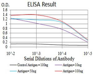
Figure 1: Black line: Control Antigen (100 ng); Purple line: Antigen(10ng); Blue line: Antigen (50 ng); Red line: Antigen (100 ng);
Western Blot

Figure 2:Western blot analysis using PRDM5 mAb against human PRDM5 (AA: 1-100) recombinant protein. (Expected MW is 37.6 kDa)
Western Blot
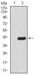
Figure 3:Western blot analysis using PRDM5 mAb against HEK293 (1) and PRDM5 (AA: 1-100)-hIgGFc transfected HEK293 (2) cell lysate.
Western Blot

Figure 4:Western blot analysis using PRDM5 mouse mAb against HL-60 (1), NIH/3T3 (2), SW480 (3), and HEK293 (4) cell lysate.
Immunofluorescence analysis

Figure 5:Immunofluorescence analysis of MCF-7 cells using PRDM5 mouse mAb (green). Blue: DRAQ5 fluorescent DNA dye. Red: Actin filaments have been labeled with Alexa Fluor- 555 phalloidin. Secondary antibody from Fisher (Cat#: 35503)
Flow cytometric

Figure 6:Flow cytometric analysis of HeLa cells using PRDM5 mouse mAb (green) and negative control (red).
For Research Use Only. Not for use in diagnostic procedures.