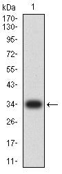
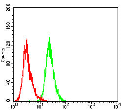
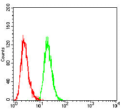
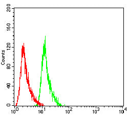
PMS2 Primary Antibody

Item Information
Catalog #
Size
Price
Description

The protein encoded by this gene is a key component of the mismatch repair system that functions to correct DNA mismatches and small insertions and deletions that can occur during DNA replication and homologous recombination. This protein forms heterodimers with the gene product of the mutL homolog 1 (MLH1) gene to form the MutL-alpha heterodimer. The MutL-alpha heterodimer possesses an endonucleolytic activity that is activated following recognition of mismatches and insertion/deletion loops by the MutS-alpha and MutS-beta heterodimers, and is necessary for removal of the mismatched DNA. There is a DQHA(X)2E(X)4E motif found at the C-terminus of the protein encoded by this gene that forms part of the active site of the nuclease. Mutations in this gene have been associated with hereditary nonpolyposis colorectal cancer (HNPCC; also known as Lynch syndrome) and Turcot syndrome.

Product Overview
Entrez GenelD
5395
Aliases
MLH4; PMSL2; HNPCC4; PMS2CL
Clone#
6G12E1
Host / Isotype
Mouse / Mouse IgG1
Immunogen
Purified recombinant fragment of human PMS2 (AA: (431-580)) expressed in E. Coli.
Formulation
Purified antibody in PBS with 0.05% sodium azide
Storage
4°C; -20°C for long term storage
Product Applications
WB (Western Blot)
1/500 - 1/2000
IHC_P(Immunohistochemistry)
1/200-1/1000
FCM (Flow Cytometry)
1/200-1/400
ELISA
1/10000
References
1,Cancer Epidemiol Biomarkers Prev. 2019 Jun;28(6):1010-1014.2,Hum Mutat. 2019 Jul;40(7):904-907.
Product Image
ELISA
Figure 1: Black line: Control Antigen (100 ng);Purple line: Antigen (10ng); Blue line: Antigen (50 ng); Red line: Antigen (100 ng)
WESTERN BLOT
Figure 2: Western blot analysis using PMS2 mAb against human PMS2 (AA: 431-580) recombinant protein. (Expected MW is 35 kDa)
WESTERN BLOT
Figure 3: Western blot analysis using PMS2 mAb against HEK293-6e (1) and PMS2 (AA: 431-580)-hIgGFc transfected HEK293-6e (2) cell lysate.
FLOW CYTOMETRY
Figure 4: Flow cytometric analysis of A431 cells using PMS2 mouse mAb (green) and negative control (red).
FLOW CYTOMETRY
Figure 5: Flow cytometric analysis of Hepg2 cells using PMS2 mouse mAb (green) and negative control (red).
FLOW CYTOMETRY
Figure 6: Flow cytometric analysis of NIH3T3 cells using PMS2 mouse mAb (green) and negative control (red).
IMMUNOHISTOCHEMISTRY
Figure 7: Immunohistochemical analysis of paraffin-embedded bladder cancer tissues using PMS2 mouse mAb with DAB staining.
IMMUNOHISTOCHEMISTRY
Figure 8: Immunohistochemical analysis of paraffin-embedded rectal cancer tissues using PMS2 mouse mAb with DAB staining.
For Research Use Only. Not for use in diagnostic procedures.