
Pirh2 Primary Antibody
Item Information
Catalog #
Size
Price
Description
Pirh 2 (P53 induced RING-H2 protein), also known as RCHY1, it forms dimers through its N- and C-terminus in cells. The Pirh2 has ubiquitin-protein ligase activity and it binds with p53 and promotes the ubiquitin-mediated proteosomal degradation of p53. The Pirh2 is oncogenic because loss of p53 function contributes directly to malignant tumor development. Pirh2 expression decreases the level of p53, and a decrease of endogenous Pirh2 expression increases p53 levels. Pirh2 is therefore considered, together with MDM2, to act as a negative regulator of p53 function.
Product Overview
Entrez GenelD
25898
Aliases
ARNIP; CHIMP; RNF199; RCHY1
Clone#
1H10
Host / Isotype
Mouse / IgG1
Species Reactivity
Human, Rat
Immunogen
Purified recombinant fragment of human Pirh2 expressed in E. Coli.
Formulation
Ascitic fluid containing 0.03% sodium azide.
Storage
Store at 4°C short term. Aliquot and store at -20°C long term. Avoid freeze/thaw cycles.
Product Applications
WB (Western Blot)
1/500 - 1/2000
IHC_P(Immunohistochemistry)
1/200 - 1/1000
ICC (Immunocytochemistry)
1/200 - 1/1000
FCM (Flow Cytometry)
1/200 - 1/400
ELISA
1/10000
References
1. Biochem Biophys Res Commun. 2007 Dec 14;364(2):344-50.
2. J Natl Cancer Inst. 2004 Nov 17;96(22):1718-21.
3. Exp Cell Res. 2006 Oct 15;312(17):3370-8.
4. Cell. 2003 Mar 21;112(6):779-91.
2. J Natl Cancer Inst. 2004 Nov 17;96(22):1718-21.
3. Exp Cell Res. 2006 Oct 15;312(17):3370-8.
4. Cell. 2003 Mar 21;112(6):779-91.
Product Image
Western Blot
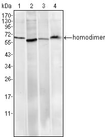
Figure 1: Western blot analysis using Pirh2 mouse mAb against Hela (1), A549 (2), MCF-7 (3) and PC-12 (4) cell lysate.
Immunohistochemical analysis

Figure 2: Immunohistochemical analysis of paraffin-embedded human Tonsil tissues using anti-Pirh2 mouse mAb
Flow cytometric

Figure 3: Flow cytometric analysis of PC-12 cells using anti-Pirh2 mAb (blue) and negative control (red).
Immunofluorescence analysis

Figure 4: Immunofluorescence analysis of Hela cells using Pirh2 mouse mAb (green). Red: Actin filaments have been labeled with Alexa Fluor-555 phalloidin.
For Research Use Only. Not for use in diagnostic procedures.