
PDX1 Primary Antibody
Item Information
Catalog #
Size
Price
Description
The protein encoded by this gene is a transcriptional activator of several genes, including insulin, somatostatin, glucokinase, islet amyloid polypeptide, and glucose transporter type 2. The encoded nuclear protein is involved in the early development of the pancreas and plays a major role in glucose-dependent regulation of insulin gene expression. Defects in this gene are a cause of pancreatic agenesis, which can lead to early-onset insulin-dependent diabetes mellitus (NIDDM), as well as maturity onset diabetes of the young type 4 (MODY4).
Product Overview
Entrez GenelD
3651
Aliases
GSF; IPF1; IUF1; IDX-1; MODY4; PDX-1; STF-1
Clone#
5A5
Host / Isotype
Mouse / IgG1
Species Reactivity
Human
Immunogen
Purified recombinant fragment of human PDX1 expressed in E. Coli.
Formulation
Purified antibody in PBS with 0.05% sodium azide
Storage
Store at 4°C short term. Aliquot and store at -20°C long term. Avoid freeze/thaw cycles.
Product Applications
WB (Western Blot)
1/500 - 1/2000
FCM (Flow Cytometry)
1/200 - 1/400
ELISA
1/10000
References
J Biol Chem. 2009 Dec 25;284(52):36482-90.
Pancreatology. 2009;9(1-2):116-26.
Pancreatology. 2009;9(1-2):116-26.
Product Image
Western Blot

Figure 1: Western blot analysis using PDX1 mAb against human PDX1 (AA: 39-283) recombinant protein. (Expected MW is 52 kDa)
Western Blot
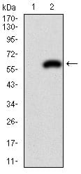
Figure 2: Western blot analysis using PDX1 mAb against HEK293 (1) and PDX1 (AA: 39-283)-hIgGFc transfected HEK293 (2) cell lysate.
Flow cytometric

Figure 3: Flow cytometric analysis of Jurkat cells using PDX1 mouse mAb (green) and negative control (red).
Elisa

Black line: Control Antigen (100 ng); Purple line: Antigen(10ng); Blue line: Antigen (50 ng); Red line: Antigen (100 ng);
For Research Use Only. Not for use in diagnostic procedures.