
MITF Primary Antibody
Item Information
Catalog #
Size
Price
Description
This gene encodes a transcription factor that contains both basic helix-loop-helix and leucine zipper structural features. It regulates the differentiation and development of melanocytes retinal pigment epithelium and is also responsible for pigment cell-specific transcription of the melanogenesis enzyme genes. Heterozygous mutations in the this gene cause auditory-pigmentary syndromes, such as Waardenburg syndrome type 2 and Tietz syndrome. Alternatively spliced transcript variants encoding different isoforms have been identified.
Product Overview
Entrez GenelD
4286
Aliases
MI; WS2; CMM8; WS2A; bHLHe32
Clone#
3A2E2
Host / Isotype
Mouse / IgG1
Species Reactivity
Human
Immunogen
Purified recombinant fragment of human MITF (AA: 1-114) expressed in E. Coli.
Formulation
Purified antibody in PBS with 0.05% sodium azide
Storage
Store at 4°C short term. Aliquot and store at -20°C long term. Avoid freeze/thaw cycles.
Product Applications
WB (Western Blot)
1/500 - 1/2000
FCM (Flow Cytometry)
1/200 - 1/400
ELISA
1/10000
References
1.Cell Mol Life Sci. 2015 Apr;72(7):1249-60.
2.Int J Clin Exp Pathol. 2013 Jul 15;6(8):1658-64.
2.Int J Clin Exp Pathol. 2013 Jul 15;6(8):1658-64.
Product Image
Elisa
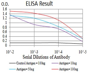
Figure 1: Black line: Control Antigen (100 ng);Purple line: Antigen (10ng); Blue line: Antigen (50 ng); Red line:Antigen (100 ng)
Western Blot

Figure 2:Western blot analysis using MITF mAb against human MITF (AA: 1-114) recombinant protein. (Expected MW is 38.9 kDa)
Western Blot
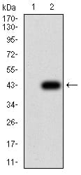
Figure 3:Western blot analysis using MITF mAb against HEK293 (1) and MITF (AA: 1-114)-hIgGFc transfected HEK293 (2) cell lysate.
Flow cytometric
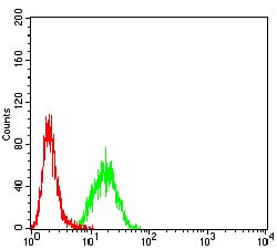
Figure 4:Flow cytometric analysis of Hela cells using MITF mouse mAb (green) and negative control (red).
For Research Use Only. Not for use in diagnostic procedures.