
MEF2C Primary Antibody
Item Information
Catalog #
Size
Price
Description
This locus encodes a member of the MADS box transcription enhancer factor 2 (MEF2) family of proteins, which play a role in myogenesis. The encoded protein, MEF2 polypeptide C, has both trans-activating and DNA binding activities. This protein may play a role in maintaining the differentiated state of muscle cells. Mutations and deletions at this locus have been associated with severe mental retardation, stereotypic movements, epilepsy, and cerebral malformation. Alternatively spliced transcript variants have been described.
Product Overview
Entrez GenelD
4208
Aliases
DEL5q14.3; C5DELq14.3
Clone#
6H2G2
Host / Isotype
Mouse / IgG1
Species Reactivity
Human, Mouse
Immunogen
Purified recombinant fragment of human MEF2C (AA: 1-125) expressed in E. Coli.
Formulation
Purified antibody in PBS with 0.05% sodium azide
Storage
Store at 4°C short term. Aliquot and store at -20°C long term. Avoid freeze/thaw cycles.
Product Applications
WB (Western Blot)
1/500 - 1/2000
IHC_P(Immunohistochemistry)
1/200 - 1/1000
FCM (Flow Cytometry)
1/200 - 1/400
ELISA
1/10000
References
1.PLoS One. 2011;6(11):e27165.
2.J Biol Chem. 2011 Aug 26;286(34):30071-86.
2.J Biol Chem. 2011 Aug 26;286(34):30071-86.
Product Image
Western Blot

Figure 1: Western blot analysis using MEF2C mAb against human MEF2C recombinant protein. (Expected MW is 40 kDa)
Western Blot

Figure 2: Western blot analysis using MEF2C mAb against HEK293 (1) and MEF2C (AA: 1-125)-hIgGFc transfected HEK293 (2) cell lysate.
Western Blot

Figure 3: Western blot analysis using MEF2C mouse mAb against NIH3T3 (1) and 3T3-L1 (2) cell lysate.
Flow cytometric
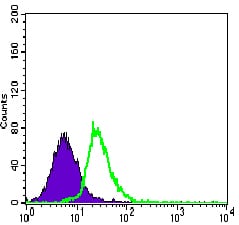
Figure 4: Flow cytometric analysis of HeLa cells using MEF2C mouse mAb (green) and negative control (purple).
Immunohistochemical analysis

Figure 5: Immunohistochemical analysis of paraffin-embedded colon cancer tissues using MEF2C mouse mAb with DAB staining.
Immunohistochemical analysis

Figure 6: Immunohistochemical analysis of paraffin-embedded esophageal cancer tissues using MEF2C mouse mAb with DAB staining.
Elisa

Black line: Control Antigen (100 ng); Purple line: Antigen(10ng); Blue line: Antigen (50 ng); Red line: Antigen (100 ng);
For Research Use Only. Not for use in diagnostic procedures.