
MB Primary Antibody
Item Information
Catalog #
Size
Price
Description
This gene encodes a member of the globin superfamily and is expressed in skeletal and cardiac muscles. The encoded protein is a haemoprotein contributing to intracellular oxygen storage and transcellular facilitated diffusion of oxygen. At least three alternatively spliced transcript variants encoding the same protein have been reported.
Product Overview
Entrez GenelD
4151
Aliases
PVALB
Clone#
6B9D2
Host / Isotype
Mouse / IgG2a
Species Reactivity
Human
Immunogen
Purified recombinant fragment of human MB (AA: 34-126) expressed in E. Coli.
Formulation
Purified antibody in PBS with 0.05% sodium azide.
Storage
Store at 4°C short term. Aliquot and store at -20°C long term. Avoid freeze/thaw cycles.
Product Applications
WB (Western Blot)
1/500 - 1/2000
IHC_P(Immunohistochemistry)
1/200 - 1/1000
ICC (Immunocytochemistry)
1/200 - 1/1000
ELISA
1/10000
References
1. Lung Cancer. 2011 Dec;74(3):411-8.
2. Br J Cancer. 2010 Jun 8;102(12):1736-45.
2. Br J Cancer. 2010 Jun 8;102(12):1736-45.
Product Image
Western Blot

Figure 1: Western blot analysis using MB mAb against human MB (AA: 34-126) recombinant protein. (Expected MW is 36.3 kDa)
Western Blot
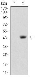
Figure 2: Western blot analysis using MB mAb against HEK293 (1) and MB (AA: 34-126)-hIgGFc transfected HEK293 (2) cell lysate.
Immunofluorescence analysis

Figure 3: Immunofluorescence analysis of Hela cells using MB mouse mAb (green). Blue: DRAQ5 fluorescent DNA dye. Red: Actin filaments have been labeled with Alexa Fluor-555 phalloidin. Secondary antibody from Fisher (Cat#: 35503)
Immunohistochemical analysis
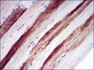
Figure 4: Immunohistochemical analysis of paraffin-embedded muscle tissues using MB mouse mAb with DAB staining.
Elisa

Black line: Control Antigen (100 ng); Purple line: Antigen(10ng); Blue line: Antigen (50 ng); Red line: Antigen (100 ng);
For Research Use Only. Not for use in diagnostic procedures.