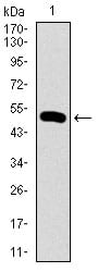
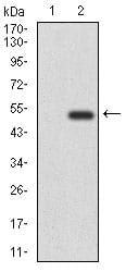
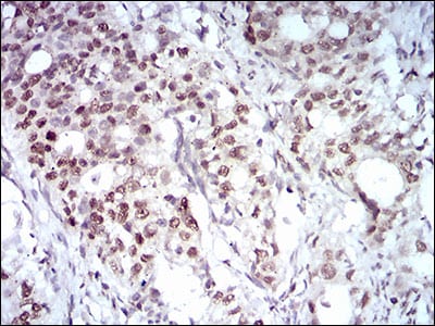
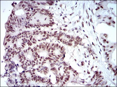
MAPK7 Primary Antibody

Item Information
Catalog #
Size
Price
Description

The protein encoded by this gene is a member of the MAP kinase family. MAP kinases act as an integration point for multiple biochemical signals, and are involved in a wide variety of cellular processes such as proliferation, differentiation, transcription regulation and development. This kinase is specifically activated by mitogen-activated protein kinase kinase 5 (MAP2K5/MEK5). It is involved in the downstream signaling processes of various receptor molecules including receptor type kinases, and G protein-coupled receptors. In response to extracelluar signals, this kinase translocates to cell nucleus, where it regulates gene expression by phosphorylating, and activating different transcription factors. Four alternatively spliced transcript variants of this gene encoding two distinct isoforms have been reported.

Product Overview
Entrez GenelD
5598
Aliases
BMK1; ERK4; ERK5; PRKM7
Clone#
8H1D5
Host / Isotype
Mouse / IgG1
Species Reactivity
Human
Immunogen
Purified recombinant fragment of human MAPK7 (AA: 411-556) expressed in E. Coli.
Formulation
Purified antibody from tissue culture in PBS with 0.05% sodium azide
Storage
4°C; -20°C for long term storage
Product Applications
WB (Western Blot)
1/500 - 1/2000
IHC_P(Immunohistochemistry)
1/200 - 1/1000
ELISA
1/10000
References
1. Br J Cancer. 2011 Feb 15;104(4):664-72.
2. PLoS One. 2009;4(5):e5565.
Product Image
Western Blot
Figure 1: Western blot analysis using MAPK7 mAb against human MAPK7 (AA: 411-556) recombinant protein. (Expected MW is 41.8 kDa)
Western Blot
Figure 2: Western blot analysis using MAPK7 mAb against HEK293 (1) and MAPK7 (AA: 411-556)-hIgGFc transfected HEK293 (2) cell lysate.
Immunohistochemical analysis
Figure 3: Immunohistochemical analysis of paraffin-embedded cervical cancer tissues using MAPK7 mouse mAb with DAB staining.
Immunohistochemical analysis
Figure 4: Immunohistochemical analysis of paraffin-embedded colon cancer tissues using MAPK7 mouse mAb with DAB staining.
Elisa
Black line: Control Antigen (100 ng); Purple line: Antigen(10ng); Blue line: Antigen (50 ng); Red line: Antigen (100 ng);
For Research Use Only. Not for use in diagnostic procedures.