
MAP2K7 Primary Antibody
Item Information
Catalog #
Size
Price
Description
The protein encoded by this gene is a dual specificity protein kinase that belongs to the MAP kinase kinase family. This kinase specifically activates MAPK8/JNK1 and MAPK9/JNK2, and this kinase itself is phosphorylated and activated by MAP kinase kinase kinases including MAP3K1/MEKK1, MAP3K2/MEKK2,MAP3K3/MEKK5, and MAP4K2/GCK. This kinase is involved in the signal transduction mediating the cell responses to proinflammatory cytokines, and environmental stresses. Multiple alternatively spliced transcript variants encoding distinct isoforms have been found, but only one transcript variant has been supported and defined.
Product Overview
Entrez GenelD
5609
Aliases
MKK7; Jnkk2; MAPKK7; PRKMK7
Clone#
4E5
Host / Isotype
Mouse / IgG1
Species Reactivity
Human
Immunogen
Purified recombinant fragment of human MAP2K7 expressed in E. Coli.
Formulation
Ascitic fluid containing 0.03% sodium azide.
Storage
Store at 4°C short term. Aliquot and store at -20°C long term. Avoid freeze/thaw cycles.
Product Applications
WB (Western Blot)
1/500 - 1/2000
IHC_P(Immunohistochemistry)
1/200 - 1/1000
ICC (Immunocytochemistry)
1/200 - 1/1000
FCM (Flow Cytometry)
1/200 - 1/400
ELISA
1/10000
References
1. Biochem J. 2010 Mar 29;427(2):237-45.
2. J Immunol. 2008 Sep 1;181(5):3252-8.
2. J Immunol. 2008 Sep 1;181(5):3252-8.
Product Image
Western Blot
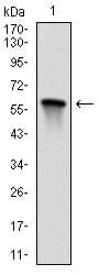
Figure 1: Western blot analysis using MAP2K7 mAb against human MAP2K7 (AA: 7-178) recombinant protein. (Expected MW is 45.1 kDa)
Immunohistochemical analysis

Figure 2: Immunohistochemical analysis of paraffin-embedded lung cancer tissues using MAP2K7 mouse mAb with DAB staining.
Immunohistochemical analysis

Figure 3: Immunohistochemical analysis of paraffin-embedded muscle tissues using MAP2K7 mouse mAb with DAB staining.
Immunofluorescence analysis

Figure 4: Immunofluorescence analysis of Hela cells using MAP2K7 mouse mAb (green). Red: Actin filaments have been labeled with Alexa Fluor-555 phalloidin.
Flow cytometric

Figure 5: Flow cytometric analysis of Hela cells using MAP2K7 mouse mAb (green) and negative control (red).
Elisa

Black line: Control Antigen (100 ng); Purple line: Antigen(10ng); Blue line: Antigen (50 ng); Red line: Antigen (100 ng);
For Research Use Only. Not for use in diagnostic procedures.