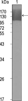
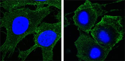
KDR Primary Antibody

Item Information
Catalog #
Size
Price
Description

KDR has also been designated as VEFR-2 (Vascular endothelial growth factor receptor 2), CD309 (cluster of differentiation 309) and Flk1 (fetal liver kinase 1). Vascular endothelial growth factor (VEGF) is a major growth factor for endothelial cells. KDR is one of the two receptors of the VEGF. This receptor, known as kinase insert domain receptor, is a type III receptor tyrosine kinase. It functions as the main mediator of VEGF-induced endothelial proliferation, survival, migration, tubular morphogenesis and sprouting. The signalling and trafficking of this receptor are regulated by multiple factors, including Rab GTPase, P2Y purine nucleotide receptor, integrin alphaVbeta3, T-cell protein tyrosine phosphatase, etc.. Mutations of this gene are implicated in infantile capillary hemangiomas.

Product Overview
Entrez GenelD
3791
Aliases
FLK1; CD309; VEGFR; VEGFR2
Clone#
4B4
Host / Isotype
Mouse / IgG1
Species Reactivity
Human
Immunogen
Purified recombinant extracellular fragment of human KDR (aa20-764) fused with hIgGFc tag expressed in HEK293 cells.
Formulation
Ascitic fluid containing 0.03% sodium azide.
Storage
4°C; -20°C for long term storage
Product Applications
WB (Western Blot)
1/500 - 1/2000
ICC (Immunocytochemistry)
1/200 - 1/1000
FCM (Flow Cytometry)
1/200 - 1/400
ELISA
1/10000
References
1. Blood. 2004 Aug 1;104(3):788-94.
2. FEBS Lett. 2002 Feb 13;512(1-3):107-10.
3. EMBO J. 2001 Jun 1;20(11):2768-78.
Product Image
Western Blot
Figure 1: Western blot analysis using KDR mouse mAb against extracellular domain of human KDR (aa20-764).
Immunofluorescence analysis
Figure 2: Confocal Immunofluorescence analysis of Hela (left) and HepG2 (right) cells using KDR mouse mAb (green). Blue: DRAQ5 fluorescent DNA dye.
Flow cytometric
Figure 3: Flow cytometric analysis of HepG2 cells using KDR mouse mAb (green) and negative control (purple).
For Research Use Only. Not for use in diagnostic procedures.