
IRAK3 Primary Antibody
Item Information
Catalog #
Size
Price
Description
This gene encodes a member of the interleukin-1 receptor-associated kinase protein family. Members of this family are essential components of the Toll/IL-R immune signal transduction pathways. This protein is primarily expressed in monocytes and macrophages and functions as a negative regulator of Toll-like receptor signaling. Mutations in this gene are associated with a susceptibility to asthma. Alternate splicing results in multiple transcript variants.
Product Overview
Entrez GenelD
11213
Aliases
ASRT5; IRAKM
Clone#
6G9G2
Host / Isotype
Mouse / IgG1
Species Reactivity
Human
Immunogen
Purified recombinant fragment of human IRAK3 (AA: 454-596) expressed in E. Coli.
Formulation
Purified antibody from tissue culture in PBS with 0.05% sodium azide
Storage
Store at 4°C short term. Aliquot and store at -20°C long term. Avoid freeze/thaw cycles.
Product Applications
WB (Western Blot)
1/500 - 1/2000
IHC_P(Immunohistochemistry)
1/200 - 1/1000
ICC (Immunocytochemistry)
1/200 - 1/1000
FCM (Flow Cytometry)
1/200 - 1/400
ELISA
1/10000
References
1.PLoS One. 2012;7(1):e30414.
2.Am J Respir Cell Mol Biol. 2011 Oct;45(4):740-5.
2.Am J Respir Cell Mol Biol. 2011 Oct;45(4):740-5.
Product Image
Elisa
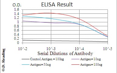
Figure 1: Black line: Control Antigen (100 ng); Purple line: Antigen(10ng); Blue line: Antigen (50 ng); Red line: Antigen (100 ng);
Western Blot
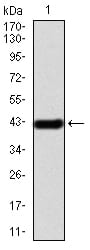
Figure 2:Western blot analysis using IRAK3 mAb against human IRAK3 (AA: 454-596) recombinant protein. (Expected MW is 42.3 kDa)
Western Blot
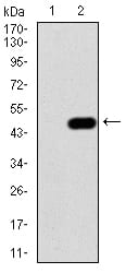
Figure 3:Western blot analysis using IRAK3 mAb against HEK293 (1) and IRAK3 (AA: 454-596)-hIgGFc transfected HEK293 (2) cell lysate.
Immunofluorescence analysis
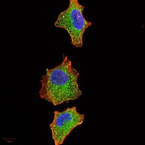
Figure 4:Immunofluorescence analysis of A549 cells using IRAK3 mouse mAb (green). Blue: DRAQ5 fluorescent DNA dye. Red: Actin filaments have been labeled with Alexa Fluor- 555 phalloidin. Secondary antibody from Fisher (Cat#: 35503)
Immunofluorescence analysis
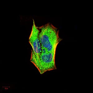
Figure 5:Immunofluorescence analysis of Hela cells using IRAK3 mouse mAb (green). Blue: DRAQ5 fluorescent DNA dye. Red: Actin filaments have been labeled with Alexa Fluor- 555 phalloidin. Secondary antibody from Fisher (Cat#: 35503)
Flow cytometric

Figure 6:Flow cytometric analysis of HepG2 cells using IRAK3 mouse mAb (green) and negative control (red).
Immunohistochemical analysis
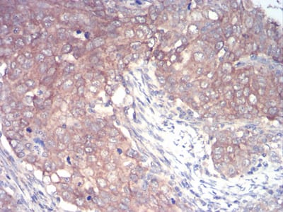
Figure 7:Immunohistochemical analysis of paraffin-embedded cervical cancer tissues using IRAK3 mouse mAb with DAB staining.
Immunohistochemical analysis
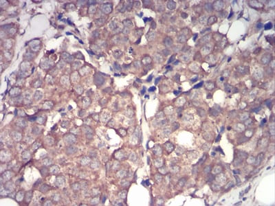
Figure 8:Immunohistochemical analysis of paraffin-embedded breast cancer tissues using IRAK3 mouse mAb with DAB staining.
For Research Use Only. Not for use in diagnostic procedures.