
Mouse Monoclonal Antibody to IL1B
Item Information
Catalog #
Size
Price
Description
The protein encoded by this gene is a member of the interleukin 1 cytokine family. This cytokine is produced by activated macrophages as a proprotein, which is proteolytically processed to its active form by caspase 1. This cytokine is an important mediator of the inflammatory response, and is involved in a variety of cellular activities, including cell proliferation, differentiation, and apoptosis. The induction of cyclooxygenase2 by this cytokine in the central nervous system is found to contribute to inflammatory pain hypersensitivity. This gene and eight other interleukin 1 family genes form a cytokine gene cluster on chromosome 2.
Product Overview
Entrez GenelD
3553
Aliases
IL-1; IL1F2; IL1beta; IL1-BETA
Clone#
5C3B7
Host / Isotype
Mouse / IgG1
Immunogen
Purified recombinant fragment of human IL1B (AA: 117-269) expressed in E. Coli.
Formulation
Purified antibody in PBS with 0.05% sodium azide
Storage
Store at 4°C short term. Aliquot and store at -20°C long term. Avoid freeze/thaw cycles.
Product Applications
WB (Western Blot)
1/500 - 1/2000
ICC (Immunocytochemistry)
1/200 - 1/1000
FCM (Flow Cytometry)
1/200 - 1/400
ELISA
1/10000
References
1,Zhonghua Jie He He Hu Xi Za Zhi. 2020 May 12;43(5):444-449.2,Medicine (Baltimore). 2020 Jul 31;99(31):e21022.
Product Image
Elisa
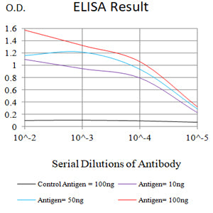
Figure 1:Black line: Control Antigen (100 ng);Purple line: Antigen (10ng); Blue line: Antigen (50 ng); Red line:Antigen (100 ng)
Western Blot

Figure 2:Western blot analysis using IL1B mAb against human IL1B (AA: 117-269) recombinant protein. (Expected MW is 43.4 kDa)
Flow cytometric analysis

Figure 3:Flow cytometric analysis of Jurkat cells using IL1B mouse mAb (green) and negative control (red).
Flow cytometric analysis

Figure 4:Flow cytometric analysis of MOLT4 cells using IL1B mouse mAb (green) and negative control (red).
Flow cytometric analysis

Figure 5:Flow cytometric analysis of THP-1 cells using IL1B mouse mAb (green) and negative control (red).
Immunofluorescence analysis

Figure 6:Immunofluorescence analysis of Hela cells using IL1B mouse mAb (green). Blue: DRAQ5 fluorescent DNA dye. Red: Actin filaments have been labeled with Alexa Fluor- 555 phalloidin. Secondary antibody from Fisher (Cat#: 35503)
For Research Use Only. Not for use in diagnostic procedures.