
IGHM Primary Antibody
Item Information
Catalog #
Size
Price
Description
Immunoglobulins (Ig) are the antigen recognition molecules of B cells. An Ig molecule is made up of 2 identical heavy chains and 2 identical light chains (see MIM 147200) joined by disulfide bonds so that each heavy chain is linked to a light chain and the 2 heavy chains are linked together. Each Ig heavy chain has an N-terminal variable (V) region containing the antigen-binding site and a C-terminal constant (C) region, encoded by an individual C region gene, that determines the isotype of the antibody and provides effector or signaling functions. The heavy chain V region is encoded by 1 each of 3 types of genes: V genes (see MIM 147070), joining (J) genes (see MIM 147010), and diversity (D) genes (see MIM 146910). The C region genes are clustered downstream of the V region genes within the heavy chain locus on chromosome 14. The IGHM gene encodes the C region of the mu heavy chain, which defines the IgM isotype. Naive B cells express the transmembrane forms of IgM and IgD (see IGHD; MIM 1471770) on their surface. During an antibody response, activated B cells can switch to the expression of individual downstream heavy chain C region genes by a process of somatic recombination known as isotype switching. In addition, secreted Ig forms that act as antibodies can be produced by alternative RNA processing of the heavy chain C region sequences. Although the membrane forms of all Ig isotypes are monomeric, secreted IgM forms pentamers, and occasionally hexamers, in plasma (summary by Janeway et al., 2005).
Product Overview
Entrez GenelD
3507
Aliases
MU; VH; AGM1
Clone#
8B9D3
Host / Isotype
Mouse / IgG1
Species Reactivity
Human
Immunogen
Purified recombinant fragment of human IGHM (AA: 310-452) expressed in E. Coli.
Formulation
Purified antibody in PBS with 0.05% sodium azide
Storage
Store at 4°C short term. Aliquot and store at -20°C long term. Avoid freeze/thaw cycles.
Product Applications
WB (Western Blot)
1/200 - 1/500
IHC_P(Immunohistochemistry)
1/200 - 1/1000
FCM (Flow Cytometry)
1/200 - 1/400
ELISA
1/10000
References
1.Cell Mol Immunol. 2014 Jan;11(1):94-104.
2.Mol Immunol. 1993 Jan;30(1):111-2.
2.Mol Immunol. 1993 Jan;30(1):111-2.
Product Image
Elisa
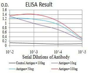
Figure 1: Black line: Control Antigen (100 ng);Purple line: Antigen (10ng); Blue line: Antigen (50 ng); Red line:Antigen (100 ng)
Western Blot

Figure 2:Western blot analysis using IGHM mAb against human IGHM (AA: 310-452) recombinant protein. (Expected MW is 41.3 kDa)
Western Blot

Figure 3:Western blot analysis using IGHM mAb against HEK293 (1) and IGHM (AA: 310-452)-hIgGFc transfected HEK293 (2) cell lysate.
Western Blot

Figure 4:Western blot analysis using IGHM mouse mAb against Raji (1) and Ramos (2) cell lysate.
Flow cytometric
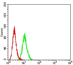
Figure 5:Flow cytometric analysis of HeLa cells using IGHM mouse mAb (green) and negative control (red).
Immunohistochemical analysis

Figure 6:Immunohistochemical analysis of paraffin-embedded endometrial cancer tissues using IGHM mouse mAb with DAB staining.
For Research Use Only. Not for use in diagnostic procedures.