
IGFBP7 Primary Antibody
Item Information
Catalog #
Size
Price
Description
This gene encodes a member of the insulin-like growth factor (IGF)-binding protein (IGFBP) family. IGFBPs bind IGFs with high affinity, and regulate IGF availability in body fluids and tissues and modulate IGF binding to its receptors. This protein binds IGF-I and IGF-II with relatively low affinity, and belongs to a subfamily of low-affinity IGFBPs. It also stimulates prostacyclin production and cell adhesion. Alternatively spliced transcript variants encoding different isoforms have been described for this gene, and one variant has been associated with retinal arterial macroaneurysm (PMID:21835307).
Product Overview
Entrez GenelD
3490
Aliases
AGM; PSF; TAF; FSTL2; IBP-7; MAC25; IGFBP-7; RAMSVPS; IGFBP-7v; IGFBPRP1
Clone#
1D9E7
Host / Isotype
Mouse / IgG1
Species Reactivity
Human
Immunogen
Purified recombinant fragment of human IGFBP7 (AA: 52-156) expressed in E. Coli.
Formulation
Purified antibody in PBS with 0.05% sodium azide
Storage
Store at 4°C short term. Aliquot and store at -20°C long term. Avoid freeze/thaw cycles.
Product Applications
WB (Western Blot)
1/500 - 1/2000
IHC_P(Immunohistochemistry)
1/200 - 1/1000
ELISA
1/10000
References
1. Sci Signal. 2012 Dec 18;5(255):ra92.
2. Cancer Biol Ther. 2012 Feb 1;13(3):148-55.
2. Cancer Biol Ther. 2012 Feb 1;13(3):148-55.
Product Image
Western Blot

Figure 1: Western blot analysis using IGFBP7 mAb against human IGFBP7 recombinant protein. (Expected MW is 36 kDa)
Western Blot

Figure 2: Western blot analysis using IGFBP7 mAb against HEK293 (1) and IGFBP7 (AA: 52-156)-hIgGFc transfected HEK293 (2) cell lysate.
Immunohistochemical analysis

Figure 3: Immunohistochemical analysis of paraffin-embedded ovarian cancer tissues using CK5 mouse mAb with DAB staining.
Immunohistochemical analysis
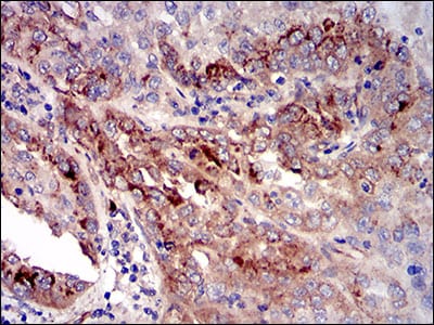
Figure 4: Immunohistochemical analysis of paraffin-embedded cervical cancer tissues using CK5 mouse mAb with DAB staining.
Elisa

Black line: Control Antigen (100 ng); Purple line: Antigen(10ng); Blue line: Antigen (50 ng); Red line: Antigen (100 ng);
For Research Use Only. Not for use in diagnostic procedures.