
HPRT1 Primary Antibody
Item Information
Catalog #
Size
Price
Description
The protein encoded by this gene is a transferase, which catalyzes conversion of hypoxanthine to inosine monophosphate and guanine to guanosine monophosphate via transfer of the 5-phosphoribosyl group from 5-phosphoribosyl 1-pyrophosphate. This enzyme plays a central role in the generation of purine nucleotides through the purine salvage pathway. Mutations in this gene result in Lesch-Nyhan syndrome or gout.
Product Overview
Entrez GenelD
3251
Aliases
HPRT; HGPRT
Clone#
5F11A7
Host / Isotype
Mouse / IgG1
Species Reactivity
Human
Immunogen
Purified recombinant fragment of human HPRT1 (AA: FULL(1-218)) expressed in E. Coli.
Formulation
Purified antibody in PBS with 0.05% sodium azide.
Storage
Store at 4°C short term. Aliquot and store at -20°C long term. Avoid freeze/thaw cycles.
Product Applications
WB (Western Blot)
1/500 - 1/2000
IHC_P(Immunohistochemistry)
1/200 - 1/1000
FCM (Flow Cytometry)
1/200 - 1/400
ELISA
1/10000
References
1. Nucleosides Nucleotides Nucleic Acids. 2011 Dec;30(12):1248-55.
2. Mol Ther. 2010 Jan;18(1):54-62.
2. Mol Ther. 2010 Jan;18(1):54-62.
Product Image
Western Blot

Figure 1: Western blot analysis using HPRT1 mAb against human HPRT1 (AA: FULL(1-218)) recombinant protein. (Expected MW is 50.5 kDa)
Western Blot
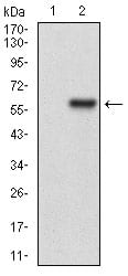
Figure 2: Western blot analysis using HPRT1 mAb against HEK293 (1) and HPRT1 (AA: FULL(1-218))-hIgGFc transfected HEK293 (2) cell lysate.
Western Blot
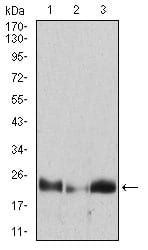
Figure 3: Western blot analysis using HPRT1 mouse mAb against Hela (1), A431 (2), A549 (3) cell lysate.
Flow cytometric

Figure 4: Flow cytometric analysis of Hela cells using HPRT1 mouse mAb (green) and negative control (red).
Immunohistochemical analysis

Figure 5: Immunohistochemical analysis of paraffin-embedded kidney tissues using HPRT1 mouse mAb with DAB staining.
Elisa
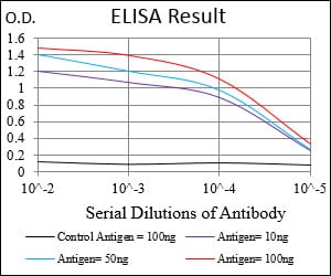
Black line: Control Antigen (100 ng); Purple line: Antigen(10ng); Blue line: Antigen (50 ng); Red line: Antigen (100 ng);
For Research Use Only. Not for use in diagnostic procedures.