
HLA-B Primary Antibody
Item Information
Catalog #
Size
Price
Description
HLA-B belongs to the HLA class I heavy chain paralogues. This class I molecule is a heterodimer consisting of a heavy chain and a light chain (beta-2 microglobulin). The heavy chain is anchored in the membrane. Class I molecules play a central role in the immune system by presenting peptides derived from the endoplasmic reticulum lumen. They are expressed in nearly all cells. The heavy chain is approximately 45 kDa and its gene contains 8 exons. Exon 1 encodes the leader peptide, exon 2 and 3 encode the alpha1 and alpha2 domains, which both bind the peptide, exon 4 encodes the alpha3 domain, exon 5 encodes the transmembrane region and exons 6 and 7 encode the cytoplasmic tail. Polymorphisms within exon 2 and exon 3 are responsible for the peptide binding specificity of each class one molecule. Typing for these polymorphisms is routinely done for bone marrow and kidney transplantation. Hundreds of HLA-B alleles have been described.
Product Overview
Entrez GenelD
3106
Aliases
AS; HLAB; Bw-47; Bw-50; SPDA1; B-4901; B-5001; HLA-Cw;
Clone#
2G7B10
Host / Isotype
Mouse / IgG1
Species Reactivity
Human
Immunogen
Purified recombinant fragment of human HLA-B (AA: 241-362) expressed in E. Coli.
Formulation
Purified antibody in PBS with 0.05% sodium azide
Storage
4°C; -20°C for long term storage
Product Applications
WB (Western Blot)
1/500 - 1/2000
IHC_P(Immunohistochemistry)
1/200 - 1/1000
ICC (Immunocytochemistry)
1/200 - 1/1000
FCM (Flow Cytometry)
1/200 - 1/400
ELISA
1/10000
References
1.Pharmacogenomics J. 2015 Oct;15(5):467-72.
2.J Immunol. 2014 Jun 1;192(11):4967-76.
2.J Immunol. 2014 Jun 1;192(11):4967-76.
Product Image
Elisa
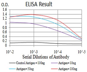
Figure 1: Black line: Control Antigen (100 ng);Purple line: Antigen (10ng); Blue line: Antigen (50 ng); Red line:Antigen (100 ng)
Western Blot

Figure 2:Western blot analysis using HLA-B mAb against human HLA-B (AA: 241-362) recombinant protein. (Expected MW is 38.4 kDa)
Western Blot

Figure 3:Western blot analysis using HLA-B mAb against HEK293 (1) and HLA-B (AA: 241-362)-hIgGFc transfected HEK293 (2) cell lysate.
Western Blot

Figure 4:Western blot analysis using HLA-B mouse mAb against Ramos (1) and A431 (2) cell lysate.
Immunofluorescence analysis

Figure 5:Immunofluorescence analysis of Hela cells using HLA-B mouse mAb. Blue: DRAQ5 fluorescent DNA dye. Red: Actin filaments have been labeled with Alexa Fluor- 555 phalloidin.
Immunofluorescence analysis

Figure 6:Immunofluorescence analysis of Hela cells using HLA-B mouse mAb (green). Blue: DRAQ5 fluorescent DNA dye. Red: Actin filaments have been labeled with Alexa Fluor- 555 phalloidin. Secondary antibody from Fisher (Cat#: 35503)
Flow cytometric

Figure 7:Flow cytometric analysis of MCF-7 cells using HLA-B mouse mAb (green) and negative control (red).
Immunohistochemical analysis

Figure 8:Immunohistochemical analysis of paraffin-embedded ovarian cancer tissues using HLA-B mouse mAb with DAB staining.
Immunohistochemical analysis
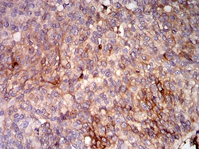
Figure 9:Immunohistochemical analysis of paraffin-embedded bladder cancer tissues using HLA-B mouse mAb with DAB staining.
For Research Use Only. Not for use in diagnostic procedures.