
HH3 Primary Antibody
Item Information
Catalog #
Size
Price
Description
Histones are basic nuclear proteins that are responsible for the nucleosome structure of the chromosomal fiber in eukaryotes. This structure consists of approximately 146 bp of DNA wrapped around a nucleosome, an octamer composed of pairs of each of the four core histones (H2A, H2B, H3, and H4). The chromatin fiber is further compacted through the interaction of a linker histone, H1, with the DNA between the nucleosomes to form higher order chromatin structures. This gene is intronless and encodes a member of the histone H3 family. Transcripts from this gene lack polyA tails; instead, they contain a palindromic termination element. This gene is found in a histone cluster on chromosome 1. This gene is one of four histone genes in the cluster that are duplicated; this record represents the centromeric copy.
Product Overview
Entrez GenelD
333932
Aliases
HIST2H3A;H3/n; H3/o
Clone#
6D3B9
Host / Isotype
Mouse / IgG1
Species Reactivity
Human, Mouse, Rat
Immunogen
Synthesized peptide fragment of human HH3 (AA: 121-136) expressed in E. Coli.
Formulation
Purified antibody from tissue culture in PBS with 0.05% sodium azide
Storage
Store at 4°C short term. Aliquot and store at -20°C long term. Avoid freeze/thaw cycles.
Product Applications
WB (Western Blot)
1/500 - 1/2000
FCM (Flow Cytometry)
1/200 - 1/400
ELISA
1/10000
References
Br J Nutr. 2010 Feb;103(3):344-51.
J Biol Chem. 2008 Feb 8;283(6):3006-10.
J Biol Chem. 2008 Feb 8;283(6):3006-10.
Product Image
Western Blot
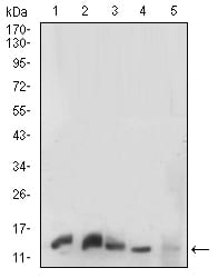
Figure 1: Western blot analysis using HH3 mouse mAb against K562 (1), C6(2),HEK293(3),PC-12(4) and NIH/3T3(5) cell lysate.
Flow cytometric
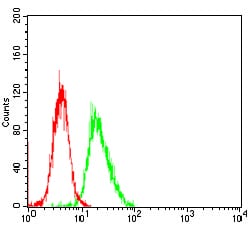
Figure 2: Flow cytometric analysis of NIH/3T3 cells using HH3 mouse mAb (green) and negative control (red).
Elisa
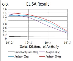
Black line: Control Antigen (100 ng); Purple line: Antigen(10ng); Blue line: Antigen (50 ng); Red line: Antigen (100 ng);
For Research Use Only. Not for use in diagnostic procedures.