
GRM5 Primary Antibody
Item Information
Catalog #
Size
Price
Description
This gene encodes a member of the G-protein coupled receptor 3 protein family. The encoded protein is a metabatropic glutamate receptor, whose signaling activates a phosphatidylinositol-calcium second messenger system. This protein may be involved in the regulation of neural network activity and synaptic plasticity. Glutamatergic neurotransmission is involved in most aspects of normal brain function and can be perturbed in many neuropathologic conditions. A pseudogene of this gene has been defined on chromosome 11. Alternative splicing results in multiple transcript variants.
Product Overview
Entrez GenelD
2915
Aliases
mGlu5; GPRC1E; MGLUR5; PPP1R86
Clone#
1A11D10
Host / Isotype
Mouse / IgG1
Species Reactivity
Human
Immunogen
Purified recombinant fragment of human GRM5 (AA: extra 458-580) expressed in E. Coli.
Formulation
Purified antibody in PBS with 0.05% sodium azide
Storage
Store at 4°C short term. Aliquot and store at -20°C long term. Avoid freeze/thaw cycles.
Product Applications
WB (Western Blot)
1/500 - 1/2000
IHC_P(Immunohistochemistry)
1/200 - 1/1000
ICC (Immunocytochemistry)
1/200 - 1/1000
ELISA
1/10000
References
1.Curr Alzheimer Res. 2014;11(7):694-705.
2.Int J Neuropsychopharmacol. 2014 Dec;17(12):1915-22.
2.Int J Neuropsychopharmacol. 2014 Dec;17(12):1915-22.
Product Image
Elisa

Figure 1: Black line: Control Antigen (100 ng);Purple line: Antigen (10ng); Blue line: Antigen (50 ng); Red line:Antigen (100 ng)
Western Blot
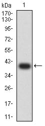
Figure 2:Western blot analysis using GRM5 mAb against human GRM5 (AA: extra 458-580) recombinant protein. (Expected MW is 40.1 kDa)
Western Blot

Figure 3:Western blot analysis using GRM5 mAb against HEK293 (1) and GRM5 (AA: extra 458-580)-hIgGFc transfected HEK293 (2) cell lysate.
Immunofluorescence analysis

Figure 4:Immunofluorescence analysis of Hela cells using GRM5 mouse mAb (green). Blue: DRAQ5 fluorescent DNA dye. Red: Actin filaments have been labeled with Alexa Fluor- 555 phalloidin. Secondary antibody from Fisher (Cat#: 35503)
Immunohistochemical analysis

Figure 5:Immunohistochemical analysis of paraffin-embedded bladder cancer tissues using GRM5 mouse mAb with DAB staining.
Immunohistochemical analysis

Figure 6:Immunohistochemical analysis of paraffin-embedded stomach cancer tissues using GRM5 mouse mAb with DAB staining.
For Research Use Only. Not for use in diagnostic procedures.