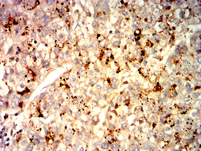
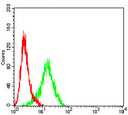
GPC3 Primary Antibody

Item Information
Catalog #
Size
Price
Description

Cell surface heparan sulfate proteoglycans are composed of a membrane-associated protein core substituted with a variable number of heparan sulfate chains. Members of the glypican-related integral membrane proteoglycan family (GRIPS) contain a core protein anchored to the cytoplasmic membrane via a glycosyl phosphatidylinositol linkage. These proteins may play a role in the control of cell division and growth regulation. The protein encoded by this gene can bind to and inhibit the dipeptidyl peptidase activity of CD26, and it can induce apoptosis in certain cell types. Deletion mutations in this gene are associated with Simpson-Golabi-Behmel syndrome, also known as Simpson dysmorphia syndrome. Alternative splicing results in multiple transcript variants.

Product Overview
Entrez GenelD
2719
Aliases
SGB; DGSX; MXR7; SDYS; SGBS; OCI-5; SGBS1; GTR2-2
Clone#
2A2C3
Host / Isotype
Mouse / Mouse IgG1
Immunogen
Purified recombinant fragment of human GPC3 (AA: 55-200) expressed in E. Coli.
Formulation
Purified antibody in PBS with 0.05% sodium azide
Storage
4°C; -20°C for long term storage
Product Applications
WB (Western Blot)
1/500 - 1/2000
IHC_P(Immunohistochemistry)
1/200-1/1000
FCM (Flow Cytometry)
1/200-1/400
ELISA
1/10000
References
1.Biomed Res Int. 2019 Nov 6;2019:2560650. 2.J Cancer Res Clin Oncol. 2018 Dec;144(12):2399-2418.
Product Image
ELISA
Figure 1: Black line: Control Antigen (100 ng);Purple line: Antigen (10ng); Blue line: Antigen (50 ng); Red line: Antigen (100 ng)
WESTERN BLOT
Figure 2: Western blot analysis using GPC3 mAb against human GPC3 (AA: 55-200) recombinant protein. (Expected MW is 29.5 kDa)
WESTERN BLOT
Figure 3: Western blot analysis using GPC3 mAb against HEK293-6e (1) and GPC3 (AA: 25-400)-hIgGFc transfected HEK293 (2) cell lysate.
WESTERN BLOT
Figure 4: Western blot analysis using GPC3 mouse mAb against HepG2 (1), BEL-7402 (2), SH-SY5Y (3), and F9 (4) cell lysate.
IMMUNOHISTOCHEMISTRY
Figure 5: Immunohistochemical analysis of paraffin-embedded liver cancer tissues using GPC3 mouse mAb with DAB staining.
FLOW CYTOMETRY
Figure 6: Flow cytometric analysis of HepG2 cells using GPC3 mouse mAb (green) and negative control (red).
For Research Use Only. Not for use in diagnostic procedures.