
GLRB Primary Antibody
Item Information
Catalog #
Size
Price
Description
This gene encodes the beta subunit of the glycine receptor, which is a pentamer composed of alpha and beta subunits. The receptor functions as a neurotransmitter-gated ion channel, which produces hyperpolarization via increased chloride conductance due to the binding of glycine to the receptor. Mutations in this gene cause startle disease, also known as hereditary hyperekplexia or congenital stiff-person syndrome, a disease characterized by muscular rigidity. Alternative splicing results in multiple transcript variants.
Product Overview
Entrez GenelD
2743
Aliases
HKPX2
Clone#
3B8A8
Host / Isotype
Mouse / IgG2b
Species Reactivity
Human
Immunogen
Purified recombinant fragment of human GLRB (AA: extra 23-160) expressed in E. Coli.
Formulation
Purified antibody in PBS with 0.05% sodium azide
Storage
Store at 4°C short term. Aliquot and store at -20°C long term. Avoid freeze/thaw cycles.
Product Applications
WB (Western Blot)
1/500 - 1/2000
ICC (Immunocytochemistry)
1/200 - 1/1000
FCM (Flow Cytometry)
1/200 - 1/400
ELISA
1/10000
References
1.Hum Mol Genet. 2013 Mar 1;22(5):927-40.
2.Clin Genet. 2012 May;81(5):479-84.
2.Clin Genet. 2012 May;81(5):479-84.
Product Image
Elisa
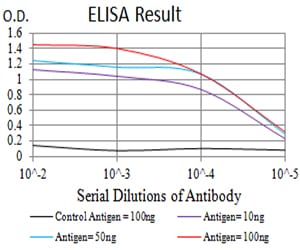
Figure 1: Black line: Control Antigen (100 ng);Purple line: Antigen (10ng); Blue line: Antigen (50 ng); Red line:Antigen (100 ng)
Western Blot
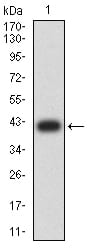
Figure 2:Western blot analysis using GLRB mAb against human GLRB (AA: extra 23-160) recombinant protein. (Expected MW is 41.8 kDa)
Western Blot
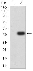
Figure 3:Western blot analysis using GLRB mAb against HEK293 (1) and GLRB (AA: extra 23-160)-hIgGFc transfected HEK293 (2) cell lysate.
Flow cytometric
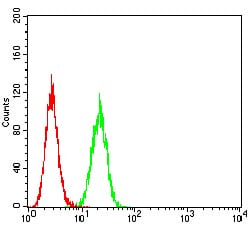
Figure 4:Flow cytometric analysis of Hela cells using GLRB mouse mAb (green) and negative control (red).
Immunofluorescence analysis
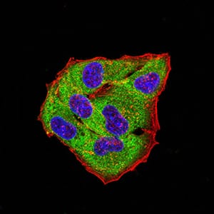
Figure 5:Immunofluorescence analysis of Hela cells using GLRB mouse mAb (green). Blue: DRAQ5 fluorescent DNA dye. Red: Actin filaments have been labeled with Alexa Fluor- 555 phalloidin. Secondary antibody from Fisher (Cat#: 35503)
For Research Use Only. Not for use in diagnostic procedures.