
EGFR Primary Antibody
Item Information
Catalog #
Size
Price
Description
The protein encoded by this gene is a transmembrane glycoprotein that is a member of the protein kinase superfamily. This protein is a receptor for members of the epidermal growth factor family. EGFR is a cell surface protein that binds to epidermal growth factor. Binding of the protein to a ligand induces receptor dimerization and tyrosine autophosphorylation and leads to cell proliferation. Mutations in this gene are associated with lung cancer. Multiple alternatively spliced transcript variants that encode different protein isoforms have been found for this gene.
Product Overview
Entrez GenelD
1956
Aliases
ERBB; HER1; mENA; ERBB1; PIG61
Clone#
5E10D3
Host / Isotype
Mouse / IgG2b
Species Reactivity
Human
Immunogen
Purified recombinant fragment of human EGFR (AA: 693-893) expressed in E. Coli.
Formulation
Purified antibody in PBS with 0.05% sodium azide
Storage
Store at 4°C short term. Aliquot and store at -20°C long term. Avoid freeze/thaw cycles.
Product Applications
WB (Western Blot)
1/500 - 1/2000
FCM (Flow Cytometry)
1/200 - 1/400
ELISA
1/10000
References
1. J Immunol. 2012 Dec 1;189(11):5230-9.
2. J Biol Chem. 2012 Oct 12;287(42):35201-11.
2. J Biol Chem. 2012 Oct 12;287(42):35201-11.
Product Image
Western Blot
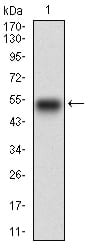
Figure 1: Western blot analysis using EGFR mAb against human EGFR recombinant protein. (Expected MW is 48.2 kDa)
Western Blot
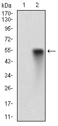
Figure 2: Western blot analysis using EGFR mAb against HEK293 (1) and EGFR (AA: 693-893)-hIgGFc transfected HEK293 (2) cell lysate.
Western Blot

Figure 3: Western blot analysis using EGFR mouse mAb against A431 (1) AND Hela (2) cell lysate.
Flow cytometric

Figure 4: Flow cytometric analysis of A431 cells using EGFR mouse mAb (green) and negative control (red).
Elisa

Black line: Control Antigen (100 ng); Purple line: Antigen(10ng); Blue line: Antigen (50 ng); Red line: Antigen (100 ng);
For Research Use Only. Not for use in diagnostic procedures.