
DSG3 Primary Antibody
Item Information
Catalog #
Size
Price
Description
Desmosomes are cell-cell junctions between epithelial, myocardial, and certain other cell types. Desmoglein 3 is a calcium-binding transmembrane glycoprotein component of desmosomes in vertebrate epithelial cells. Currently, three desmoglein subfamily members have been identified and all are members of the cadherin cell adhesion molecule superfamily. These desmoglein gene family members are located in a cluster on chromosome 18. This protein has been identified as the autoantigen of the autoimmune skin blistering disease pemphigus vulgaris.
Product Overview
Entrez GenelD
1830
Aliases
PVA; CDHF6
Clone#
6G2C11
Host / Isotype
Mouse / IgG1
Species Reactivity
Human
Immunogen
Purified recombinant fragment of human DSG3 (AA: 55-159) expressed in E. Coli.
Formulation
Purified antibody in PBS with 0.05% sodium azide.
Storage
Store at 4°C short term. Aliquot and store at -20°C long term. Avoid freeze/thaw cycles.
Product Applications
WB (Western Blot)
1/500 - 1/2000
IHC_P(Immunohistochemistry)
1/200 - 1/1000
FCM (Flow Cytometry)
1/200 - 1/400
ELISA
1/10000
References
1. J Dermatol Sci. 2012 Feb;65(2):102-9.
2. Am J Pathol. 2009 May;174(5):1629-37.
2. Am J Pathol. 2009 May;174(5):1629-37.
Product Image
Western Blot

Figure 1: Western blot analysis using DSG3 mAb against human DSG3 (AA: 55-159) recombinant protein. (Expected MW is 37.5 kDa)
Western Blot

Figure 2: Western blot analysis using DSG3 mAb against HEK293 (1) and DSG3 (AA: 55-159)-hIgGFc transfected HEK293 (2) cell lysate.
Western Blot

Figure 3: Western blot analysis using DSG3 mouse mAb against A431 cell lysate.
Flow cytometric

Figure 4: Flow cytometric analysis of A431 cells using DSG3 mouse mAb (green) and negative control (red).
Immunohistochemical analysis
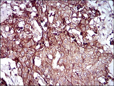
Figure 5: Immunohistochemical analysis of paraffin-embedded esophageal cancer tissues using DSG3 mouse mAb with DAB staining.
Immunohistochemical analysis

Figure 6: Immunohistochemical analysis of paraffin-embedded esophageal tissues using DSG3 mouse mAb with DAB staining.
Elisa

Black line: Control Antigen (100 ng); Purple line: Antigen(10ng); Blue line: Antigen (50 ng); Red line: Antigen (100 ng);
For Research Use Only. Not for use in diagnostic procedures.