
DLL3 Primary Antibody
Item Information
Catalog #
Size
Price
Description
This gene encodes a member of the delta protein ligand family. This family functions as Notch ligands that are characterized by a DSL domain, EGF repeats, and a transmembrane domain. Mutations in this gene cause autosomal recessive spondylocostal dysostosis 1. Two transcript variants encoding distinct isoforms have been identified for this gene.
Product Overview
Entrez GenelD
10683
Aliases
SCDO1
Clone#
1H9C2
Host / Isotype
Mouse / Mouse IgG1
Species Reactivity
Human
Immunogen
Purified recombinant fragment of human DLL3 (AA: extra 27-226) expressed in E. Coli.
Formulation
Purified antibody in PBS with 0.05% sodium azide
Storage
Store at 4°C short term. Aliquot and store at -20°C long term. Avoid freeze/thaw cycles.
Product Applications
WB (Western Blot)
1/500 - 1/2000
IHC_P(Immunohistochemistry)
1/200 - 1/1000
FCM (Flow Cytometry)
1/200 - 1/400
ELISA
1/10000
References
1,Cancer Sci . 2021 Aug;112(8):2984-2992.
2,Lung Cancer . 2019 Dec;138:102-108.
2,Lung Cancer . 2019 Dec;138:102-108.
Product Image
Elisa

Figure 1:Black line: Control Antigen (100 ng);Purple line: Antigen (10ng); Blue line: Antigen (50 ng); Red line:Antigen (100 ng)
Western Blot
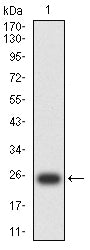
Figure 2:Western blot analysis using DLL3 mAb against human DLL3 (AA: 27-226) recombinant protein. (Expected MW is 24 kDa)
Western Blot

Figure 3:Western blot analysis using DLL3 mAb against HEK293-6e (1) and DLL3 (AA: 27-226)-hIgGFc transfected HEK293-6e (2) cell lysate.
Immunofluorescence analysis

Figure 4:Flow cytometric analysis of HEK293 cells using DLL3 mouse mAb (green) and negative control (red).
Immunohistochemical analysis

Figure 5:Immunohistochemical analysis of paraffin-embedded human brain tissues using DLL3 mouse mAb with DAB staining.
Immunohistochemical analysis

Figure 6:Immunohistochemical analysis of paraffin-embedded rectum cancer tissues using DLL3 mouse mAb with DAB staining.
For Research Use Only. Not for use in diagnostic procedures.