
CREB1 Primary Antibody
Item Information
Catalog #
Size
Price
Description
This gene encodes a transcription factor that is a member of the leucine zipper family of DNA binding proteins. This protein binds as a homodimer to the cAMP-responsive element, an octameric palindrome. The protein is phosphorylated by several protein kinases, and induces transcription of genes in response to hormonal stimulation of the cAMP pathway. Alternate splicing of this gene results in two transcript variants encoding different isoforms. (provided by RefSeq)
Product Overview
Entrez GenelD
1385
Aliases
CREB; MGC9284; CREB1
Clone#
5G3
Host / Isotype
Mouse / IgG1
Species Reactivity
Human, Mouse, Rat, Monkey
Immunogen
Purified recombinant fragment of human CREB1 expressed in E. Coli.
Formulation
Ascitic fluid containing 0.03% sodium azide.
Storage
Store at 4°C short term. Aliquot and store at -20°C long term. Avoid freeze/thaw cycles.
Product Applications
WB (Western Blot)
1/500 - 1/2000
IHC_P(Immunohistochemistry)
1/200 - 1/1000
ICC (Immunocytochemistry)
1/200 - 1/1000
ELISA
1/10000
References
1. Proc Natl Acad Sci U S A. 2008 Jul 22;105(29):10161-6.
2. FEBS Lett. 2008 Jun 11;582(13):1889-93.3
3. Am J Med Genet B Neuropsychiatr Genet. 2008 Jun 5;147B(4):500-4.
2. FEBS Lett. 2008 Jun 11;582(13):1889-93.3
3. Am J Med Genet B Neuropsychiatr Genet. 2008 Jun 5;147B(4):500-4.
Product Image
Western Blot

Figure 1: Western blot analysis using CREB1 mouse mAb against K562 (1), Jurkat (2), L1210 (3), HEK293 (4), A431 (5), Hela (6), Cos7 (7), PC-12 (8), and NIH/3T3 (9) cell lysate.
Immunohistochemical analysis
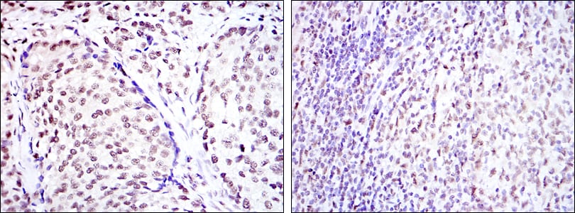
Figure 2: Immunohistochemical analysis of paraffin-embedded prostate cancer tissues (left) and submaxillary tumor tissues (right) using CREB1 mouse mAb with DAB staining.
Immunofluorescence analysis

Figure 3: Immunofluorescence analysis of Hela cells using CREB1 mouse mAb (green). Red: Actin filaments have been labeled with Alexa Fluor-555 phalloidin.
Elisa

Red: Control Antigen (100ng); Purple: Antigen (10ng); Green: Antigen (50ng); Blue: Antigen (100ng);
For Research Use Only. Not for use in diagnostic procedures.