
CPV-NS1 Primary Antibody
Item Information
Catalog #
Size
Price
Product Overview
Entrez GenelD
EF011664.1
Aliases
Canine parvovirus nonstructural protein 1
Clone#
2B5B7
Host / Isotype
Mouse / IgG1
Species Reactivity
Human, Mouse, Monkey
Immunogen
Purified recombinant fragment of Canine Parvovirus CPV-NS1 (AA: 544-668) expressed in E. Coli.
Formulation
Purified antibody in PBS with 0.05% sodium azide
Storage
4°C; -20°C for long term storage
Product Applications
WB (Western Blot)
1/500 - 1/2000
IHC_P(Immunohistochemistry)
1/200 - 1/1000
ICC (Immunocytochemistry)
1/200 - 1/1000
FCM (Flow Cytometry)
1/200 - 1/400
ELISA
1/10000
References
N
Product Image
Elisa

Figure 1:Black line: Control Antigen (100 ng);Purple line: Antigen (10ng); Blue line: Antigen (50 ng); Red line:Antigen (100 ng)
Western Blot

Figure 2:Western blot analysis using CPV-NS1 mAb against Canine CPV-NS1 (AA: 544-668) recombinant protein. (Expected MW is 40 kDa)
Western Blot
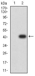
Figure 3:Western blot analysis using CPV-NS1 mAb against HEK293 (1) and CPV-NS1 (AA: 544-668)-hIgGFc transfected HEK293 (2) cell lysate.
Western Blot

Figure 4:Western blot analysis using CPV-NS1 mouse mAb against K562 (1), BCBL-1 (2), Raw264.7 (3), and COS7 (4) cell lysate.
Immunofluorescence analysis

Figure 5:Immunofluorescence analysis of Hela cells using CPV-NS1 mouse mAb (green). Blue: DRAQ5 fluorescent DNA dye. Red: Actin filaments have been labeled with Alexa Fluor- 555 phalloidin. Secondary antibody from Fisher (Cat#: 35503)
Flow cytometric

Figure 6:Flow cytometric analysis of Hela cells using CPV-NS1 mouse mAb (green) and negative control (red).
Immunohistochemical analysis

Figure 7:Immunohistochemical analysis of paraffin-embedded ovarian cancer tissues using CPV-NS1 mouse mAb with DAB staining.
Immunohistochemical analysis

Figure 8:Immunohistochemical analysis of paraffin-embedded rectum cancer tissues using CPV-NS1 mouse mAb with DAB staining.
For Research Use Only. Not for use in diagnostic procedures.