
CNN1 Primary Antibody
Item Information
Catalog #
Size
Price
Description
Calponin h1 (CN) is a differentiation marker of smooth muscle cells that has been reported to be down-regulated in the blood vessels of several human tumors.
Product Overview
Entrez GenelD
1264
Aliases
SMCC; Sm-Calp
Clone#
1H5B5
Host / Isotype
Mouse / IgG1
Species Reactivity
Human
Immunogen
Purified recombinant fragment of human CNN1 (AA: 16-165) expressed in E. Coli.
Formulation
Purified antibody from tissue culture in PBS with 0.05% sodium azide
Storage
Store at 4°C short term. Aliquot and store at -20°C long term. Avoid freeze/thaw cycles.
Product Applications
WB (Western Blot)
1/500 - 1/2000
IHC_P(Immunohistochemistry)
1/200 - 1/1000
ELISA
1/10000
References
1. Anticancer Res. 2010 Apr;30(4):1071-8.
2. Eur J Surg Oncol. 2008 May;34(5):531-7.
2. Eur J Surg Oncol. 2008 May;34(5):531-7.
Product Image
Western Blot
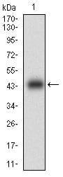
Figure 1: Western blot analysis using CNN1 mAb against human CNN1 (AA: 16-165) recombinant protein. (Expected MW is 43.1 kDa)
Western Blot
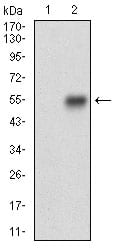
Figure 2: Western blot analysis using CNN1 mAb against HEK293 (1) and CNN1 (AA: 16-165)-hIgGFc transfected HEK293 (2) cell lysate.
Immunohistochemical analysis
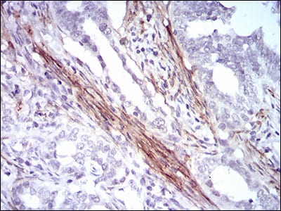
Figure 3: Immunohistochemical analysis of paraffin-embedded cervical cancer tissues using CNN1 mouse mAb with DAB staining.
Immunohistochemical analysis
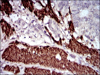
Figure 4: Immunohistochemical analysis of paraffin-embedded esophageal cancer tissues using CNN1 mouse mAb with DAB staining.
Elisa
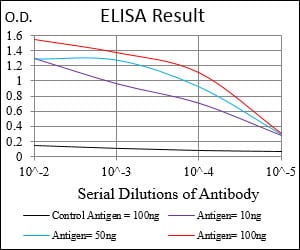
Black line: Control Antigen (100 ng); Purple line: Antigen(10ng); Blue line: Antigen (50 ng); Red line: Antigen (100 ng);
For Research Use Only. Not for use in diagnostic procedures.