
CDKN2A Primary Antibody
Item Information
Catalog #
Size
Price
Description
This gene generates several transcript variants which differ in their first exons. At least three alternatively spliced variants encoding distinct proteins have been reported, two of which encode structurally related isoforms known to function as inhibitors of CDK4 kinase. The remaining transcript includes an alternate first exon located 20 Kb upstream of the remainder of the gene; this transcript contains an alternate open reading frame (ARF) that specifies a protein which is structurally unrelated to the products of the other variants. This ARF product functions as a stabilizer of the tumor suppressor protein p53 as it can interact with, and sequester, MDM1, a protein responsible for the degradation of p53. In spite of the structural and functional differences, the CDK inhibitor isoforms and the ARF product encoded by this gene, through the regulatory roles of CDK4 and p53 in cell cycle G1 progression, share a common functionality in cell cycle G1 control. This gene is frequently mutated or deleted in a wide variety of tumors, and is known to be an important tumor suppressor gene.
Product Overview
Entrez GenelD
1029
Aliases
ARF; MLM; P14; P16; P19; CMM2; INK4; MTS1; TP16; CDK4I; CDKN2; INK4A; MTS-1; P14ARF; P19ARF; P16INK4; P16INK4A; P16-INK4A
Clone#
1D7D2A1
Host / Isotype
Mouse / IgG1
Species Reactivity
Human
Immunogen
Purified recombinant fragment of human CDKN2A (AA: 1-156) expressed in E. Coli.
Formulation
Purified antibody from tissue culture in PBS with 0.05% sodium azide
Storage
Store at 4°C short term. Aliquot and store at -20°C long term. Avoid freeze/thaw cycles.
Product Applications
WB (Western Blot)
1/500 - 1/2000
IHC_P(Immunohistochemistry)
1/200 - 1/1000
FCM (Flow Cytometry)
1/200 - 1/400
ELISA
1/10000
References
1. Clin Cancer Res. 2011 Dec 1;17(23):7413-23.
2. Appl Immunohistochem Mol Morphol. 2011 Dec;19(6):562-8.
2. Appl Immunohistochem Mol Morphol. 2011 Dec;19(6):562-8.
Product Image
Western Blot

Figure 1: Western blot analysis using CDKN2A mAb against human CDKN2A (AA: 1-156) recombinant protein. (Expected MW is 19 kDa)
Western Blot
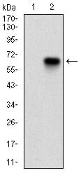
Figure 2: Western blot analysis using CDKN2A mAb against HEK293 (1) and CDKN2A (AA: 1-156)-hIgGFc transfected HEK293 (2) cell lysate.
Western Blot
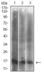
Figure 3: Western blot analysis using CDKN2A mouse mAb against Hela (1), HepG2 (2) and Hek293 (3) cell lysate.
Flow cytometric
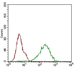
Figure 4: Flow cytometric analysis of Hela cells using CDKN2A mouse mAb (green) and negative control (red).
Immunohistochemical analysis
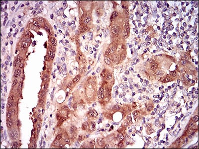
Figure 5: Immunohistochemical analysis of paraffin-embedded endometrial cancer tissues using CDKN2A mouse mAb with DAB staining.
Immunohistochemical analysis
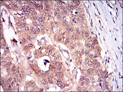
Figure 6: Immunohistochemical analysis of paraffin-embedded esophageal cancer tissues using CDKN2A mouse mAb with DAB staining.
Elisa
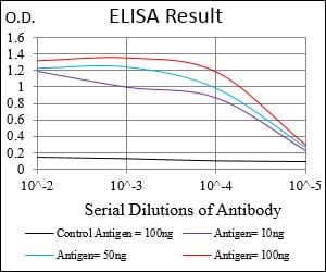
Black line: Control Antigen (100 ng); Purple line: Antigen(10ng); Blue line: Antigen (50 ng); Red line: Antigen (100 ng);
For Research Use Only. Not for use in diagnostic procedures.