
CD93 Primary Antibody
Item Information
Catalog #
Size
Price
Description
The protein encoded by this gene is a cell-surface glycoprotein and type I membrane protein that was originally identified as a myeloid cell-specific marker. The encoded protein was once thought to be a receptor for C1q, but now is thought to instead be involved in intercellular adhesion and in the clearance of apoptotic cells. The intracellular cytoplasmic tail of this protein has been found to interact with moesin, a protein known to play a role in linking transmembrane proteins to the cytoskeleton and in the remodelling of the cytoskeleton.
Product Overview
Entrez GenelD
22918
Aliases
C1QR1; C1qRP; CDw93; ECSM3; MXRA4; C1qR(P); dJ737E23.1
Clone#
1A10E10
Host / Isotype
Mouse / IgG1
Species Reactivity
Human
Immunogen
Purified recombinant fragment of human CD93 (AA: 474-535) expressed in E. Coli.
Formulation
Ascitic fluid containing 0.03% sodium azide.
Storage
Store at 4°C short term. Aliquot and store at -20°C long term. Avoid freeze/thaw cycles.
Product Applications
WB (Western Blot)
1/500 - 1/2000
IHC_P(Immunohistochemistry)
1/200 - 1/1000
ICC (Immunocytochemistry)
1/200 - 1/1000
FCM (Flow Cytometry)
1/200 - 1/400
ELISA
1/10000
References
1. PLoS One. 2012;7(12):e51647.
2. J Clin Immunol. 2010 Sep;30(5):723-33.
2. J Clin Immunol. 2010 Sep;30(5):723-33.
Product Image
Western Blot

Figure 1: Western blot analysis using CD93 mAb against human CD93 recombinant protein. (Expected MW is 31.7 kDa)
Western Blot
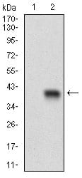
Figure 2: Western blot analysis using CD93 mAb against HEK293 (1) and CD93 (AA: 474-535)-hIgGFc transfected HEK293 (2) cell lysate.
Immunofluorescence analysis

Figure 3: Immunofluorescence analysis of Hela cells using CD93 mouse mAb (green). Blue: DRAQ5 fluorescent DNA dye.
Flow cytometric

Figure 4: Flow cytometric analysis of HepG2 cells using CD93 mouse mAb (green) and negative control (red).
Immunohistochemical analysis

Figure 5: Immunohistochemical analysis of paraffin-embedded cervical cancer tissues using CD93 mouse mAb with DAB staining.
Elisa
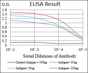
Black line: Control Antigen (100 ng); Purple line: Antigen(10ng); Blue line: Antigen (50 ng); Red line: Antigen (100 ng);
For Research Use Only. Not for use in diagnostic procedures.