
CD68 Primary Antibody
Item Information
Catalog #
Size
Price
Description
This gene encodes a 110-kD transmembrane glycoprotein that is highly expressed by human monocytes and tissue macrophages. It is a member of the lysosomal/endosomal-associated membrane glycoprotein (LAMP) family. The protein primarily localizes to lysosomes and endosomes with a smaller fraction circulating to the cell surface. It is a type I integral membrane protein with a heavily glycosylated extracellular domain and binds to tissue- and organ-specific lectins or selectins. The protein is also a member of the scavenger receptor family. Scavenger receptors typically function to clear cellular debris, promote phagocytosis, and mediate the recruitment and activation of macrophages. Alternative splicing results in multiple transcripts encoding different isoforms.
Product Overview
Entrez GenelD
968
Aliases
GP110; LAMP4; SCARD1
Clone#
3F7D3
Host / Isotype
Mouse / IgG1
Species Reactivity
Human
Immunogen
Purified recombinant fragment of human CD68 (AA: 42-155) expressed in E. Coli.
Formulation
Purified antibody from tissue culture in PBS with 0.05% sodium azide
Storage
Store at 4°C short term. Aliquot and store at -20°C long term. Avoid freeze/thaw cycles.
Product Applications
WB (Western Blot)
1/500 - 1/2000
IHC_P(Immunohistochemistry)
1/200 - 1/1000
ICC (Immunocytochemistry)
1/200 - 1/1000
FCM (Flow Cytometry)
1/200 - 1/400
ELISA
1/10000
References
1. Rom J Morphol Embryol. 2012;53(1):61-6.
2. Anticancer Res. 2009 Aug;29(8):3269-79.
2. Anticancer Res. 2009 Aug;29(8):3269-79.
Product Image
Western Blot
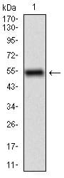
Figure 1: Western blot analysis using CD68 mAb against human CD68 (AA: 42-155) recombinant protein. (Expected MW is 37.4 kDa)
Western Blot
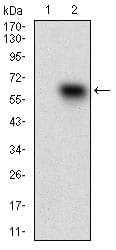
Figure 2: Western blot analysis using CD68 mAb against HEK293 (1) and CD68 (AA: 42-155)-hIgGFc transfected HEK293 (2) cell lysate.
Western Blot
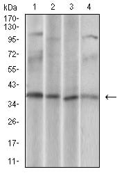
Figure 3: Western blot analysis using CD68 mouse mAb against U937 (1), Hela (2), HepG2 (3), Jurkat (4) cell lysate.
Immunofluorescence analysis
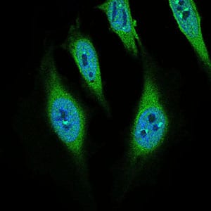
Figure 4: Immunofluorescence analysis of Hela cells using CD68 mouse mAb (green). Blue: DRAQ5 fluorescent DNA dye.
Flow cytometric
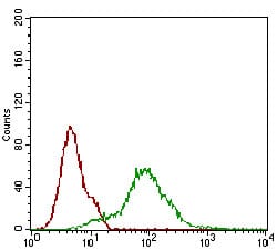
Figure 5: Flow cytometric analysis of Hela cells using CD68 mouse mAb (green) and negative control (red).
Immunohistochemical analysis
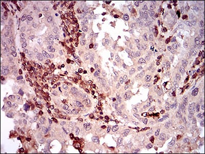
Figure 6: Immunohistochemical analysis of paraffin-embedded endometrial cancer tissues using CD68 mouse mAb with DAB staining.
Immunohistochemical analysis
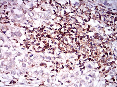
Figure 7: Immunohistochemical analysis of paraffin-embedded bladder cancer tissues using CD68 mouse mAb with DAB staining.
Elisa
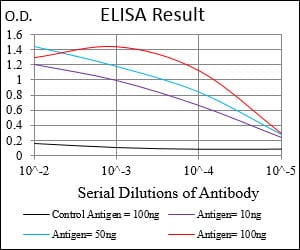
Black line: Control Antigen (100 ng); Purple line: Antigen(10ng); Blue line: Antigen (50 ng); Red line: Antigen (100 ng);
For Research Use Only. Not for use in diagnostic procedures.