
CD63 Primary Antibody
Item Information
Catalog #
Size
Price
Description
The protein encoded by this gene is a member of the transmembrane 4 superfamily, also known as the tetraspanin family. Most of these members are cell-surface proteins that are characterized by the presence of four hydrophobic domains. The proteins mediate signal transduction events that play a role in the regulation of cell development, activation, growth and motility. The encoded protein is a cell surface glycoprotein that is known to complex with integrins. It may function as a blood platelet activation marker. Deficiency of this protein is associated with Hermansky-Pudlak syndrome. Also this gene has been associated with tumor progression. Alternative splicing results in multiple transcript variants encoding different protein isoforms.
Product Overview
Entrez GenelD
967
Aliases
MLA1; ME491; LAMP-3; OMA81H; TSPAN30
Clone#
1E8D4
Host / Isotype
Mouse / IgG1
Species Reactivity
Human
Immunogen
Purified recombinant fragment of human CD63 (AA: extra 103-203) expressed in E. Coli.
Formulation
Purified antibody in PBS with 0.05% sodium azide
Storage
Store at 4°C short term. Aliquot and store at -20°C long term. Avoid freeze/thaw cycles.
Product Applications
WB (Western Blot)
1/500 - 1/2000
IHC_P(Immunohistochemistry)
1/200 - 1/1000
FCM (Flow Cytometry)
1/200 - 1/400
ELISA
1/10000
References
1.J Invest Dermatol. 2014 Dec;134(12):2947-2956.
2.Eur J Immunol. 2011 Sep;41(9):2556-61.
2.Eur J Immunol. 2011 Sep;41(9):2556-61.
Product Image
Elisa

Figure 1: Black line: Control Antigen (100 ng);Purple line: Antigen (10ng); Blue line: Antigen (50 ng); Red line:Antigen (100 ng)
Western Blot

Figure 2:Western blot analysis using CD63 mAb against human CD63 (AA: extra 103-203) recombinant protein. (Expected MW is 37.4 kDa)
Western Blot

Figure 3:Western blot analysis using CD63 mAb against HEK293 (1) and CD63 (AA: extra 103-203)-hIgGFc transfected HEK293 (2) cell lysate.
Flow cytometric
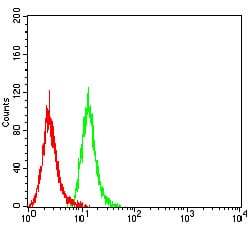
Figure 4:Flow cytometric analysis of Hela cells using CD63 mouse mAb (green) and negative control (red).
Immunohistochemical analysis

Figure 5:Immunohistochemical analysis of paraffin-embedded cervical cancer tissues using CD63 mouse mAb with DAB staining.
Immunohistochemical analysis

Figure 6:Immunohistochemical analysis of paraffin-embedded rectum cancer tissues using CD63 mouse mAb with DAB staining.
For Research Use Only. Not for use in diagnostic procedures.