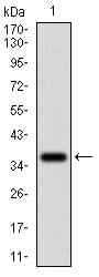
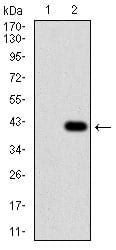
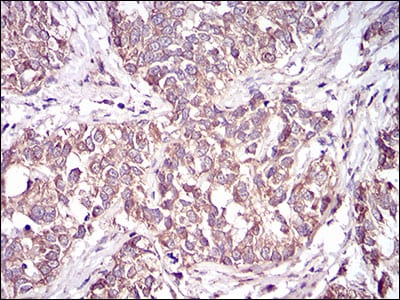
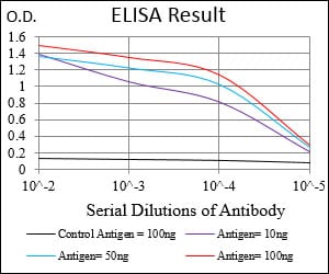
CD36 Primary Antibody

Item Information
Catalog #
Size
Price
Description

The protein encoded by this gene is the fourth major glycoprotein of the platelet surface and serves as a receptor for thrombospondin in platelets and various cell lines. Since thrombospondins are widely distributed proteins involved in a variety of adhesive processes, this protein may have important functions as a cell adhesion molecule. It binds to collagen, thrombospondin, anionic phospholipids and oxidized LDL. It directly mediates cytoadherence of Plasmodium falciparum parasitized erythrocytes and it binds long chain fatty acids and may function in the transport and/or as a regulator of fatty acid transport. Mutations in this gene cause platelet glycoprotein deficiency. Multiple alternatively spliced transcript variants encoding the same protein have been found for this gene.

Product Overview
Entrez GenelD
948
Aliases
FAT; GP4; GP3B; GPIV; CHDS7; PASIV; SCARB3; BDPLT10
Clone#
5B6F5
Host / Isotype
Mouse / IgG1
Species Reactivity
Human
Immunogen
Purified recombinant fragment of human CD36 (AA: 30-130) expressed in E. Coli.
Formulation
Purified antibody from tissue culture in PBS with 0.05% sodium azide
Storage
4°C; -20°C for long term storage
Product Applications
WB (Western Blot)
1/500 - 1/2000
IHC_P(Immunohistochemistry)
1/200 - 1/1000
ELISA
1/10000
References
1. Blood. 2011 Jun 9;117(23):6355-66.
2. J Thromb Haemost. 2011 Sep;9(9):1835-46.
Product Image
Western Blot
Figure 1: Western blot analysis using CD36 mAb against human CD36 (AA: 30-130) recombinant protein. (Expected MW is 37.5 kDa)
Western Blot
Figure 2: Western blot analysis using CD36 mAb against HEK293 (1) and CD36 (AA: 30-130)-hIgGFc transfected HEK293 (2) cell lysate.
Immunohistochemical analysis
Figure 3: Immunohistochemical analysis of paraffin-embedded bladder cancer tissues using CD36 mouse mAb with DAB staining.
Elisa
Black line: Control Antigen (100 ng); Purple line: Antigen(10ng); Blue line: Antigen (50 ng); Red line: Antigen (100 ng);
For Research Use Only. Not for use in diagnostic procedures.