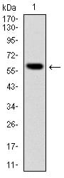
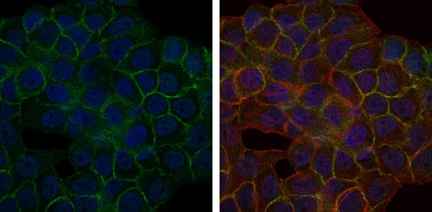
BLNK Primary Antibody

Item Information
Catalog #
Size
Price
Description

This gene encodes a cytoplasmic linker or adaptor protein that plays a critical role in B cell development. This protein bridges B cell receptor-associated kinase activation with downstream signaling pathways, thereby affecting various biological functions. The phosphorylation of five tyrosine residues is necessary for this protein to nucleate distinct signaling effectors following B cell receptor activation. Mutations in this gene cause hypoglobulinemia and absent B cells, a disease in which the pro- to pre-B-cell transition is developmentally blocked. Deficiency in this protein has also been shown in some cases of pre-B acute lymphoblastic leukemia. Alternatively spliced transcript variants encoding different isoforms have been found for this gene.

Product Overview
Entrez GenelD
29760
Aliases
AGM4; BASH; LY57; SLP65; BLNK-S; SLP-65; MGC111051
Clone#
5G9
Host / Isotype
Mouse / IgG1
Species Reactivity
Human, Mouse
Immunogen
Purified recombinant fragment of human BLNK expressed in E. Coli.
Formulation
Ascitic fluid containing 0.03% sodium azide.
Storage
4°C; -20°C for long term storage
Product Applications
WB (Western Blot)
1/500 - 1/2000
IHC_P(Immunohistochemistry)
1/200 - 1/1000
ICC (Immunocytochemistry)
1/200 - 1/1000
FCM (Flow Cytometry)
1/200 - 1/400
ELISA
1/10000
References
1. J Biol Chem. 2009 Apr 10;284(15):9804-13.
2. Cancer Sci. 2008 Dec;99(12):2444-54.
Product Image
Western Blot
Figure 1: Western blot analysis using BLNK mAb against human BLNK (AA: 34-216) recombinant protein. (Expected MW is 60 kDa)
Western Blot
Figure 2: Western blot analysis using BLNK mouse mAb against NIH/3T3 (1) and BCBL-1 (2) cell lysate.
Immunohistochemical analysis
Figure 3: Immunohistochemical analysis of paraffin-embedded human cervical cancer tissues using BLNK mouse mAb with DAB staining.
Immunofluorescence analysis
Figure 4: Immunofluorescence analysis of HepG2 cells using BLNK mouse mAb (green). Blue: DRAQ5 fluorescent DNA dye. Red: Actin filaments have been labeled with Alexa Fluor-555 phalloidin.
Flow cytometric
Figure 5: Flow cytometric analysis of NIH/3T3 cells using BLNK mouse mAb (green) and negative control (purple).
Elisa
Red: Control Antigen (100ng); Purple: Antigen (10ng); Green: Antigen (50ng); Blue: Antigen (100ng);
For Research Use Only. Not for use in diagnostic procedures.