

BCL11B Primary Antibody
Item Information
Catalog #
Size
Price
Description
This gene encodes a C2H2-type zinc finger protein and is closely related to BCL11A, a gene whose translocation may be associated with B-cell malignancies. Although the specific function of this gene has not been determined, the encoded protein is known to be a transcriptional repressor, and is regulated by the NURD nucleosome remodeling and histone deacetylase complex. Four alternatively spliced transcript variants encoding distinct isoforms have been found for this gene.
Product Overview
Entrez GenelD
64919
Aliases
ATL1; RIT1; CTIP2; IMD49; CTIP-2; ZNF856B; ATL1-beta; ATL1-alpha; ATL1-delta; ATL1-gamma; hRIT1-alpha
Clone#
1F8G8
Host / Isotype
Mouse / IgG1
Species Reactivity
Human
Immunogen
Purified recombinant fragment of human BCL11B (AA: 1-150) expressed in E. Coli.
Formulation
Purified antibody in PBS with 0.05% sodium azide
Storage
Store at 4°C short term. Aliquot and store at -20°C long term. Avoid freeze/thaw cycles.
Product Applications
WB (Western Blot)
1/500 - 1/2000
IHC_P(Immunohistochemistry)
1/200 - 1/1000
ICC (Immunocytochemistry)
1/50 - 1/250
FCM (Flow Cytometry)
1/200 - 1/400
ELISA
1/10000
References
1.J Immunol. 2014 Sep 1;193(5):2059-65.
2.PLoS One. 2013;8(1):e55147.
2.PLoS One. 2013;8(1):e55147.
Product Image
Elisa
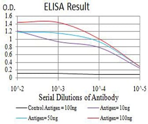
Figure 1: Black line: Control Antigen (100 ng);Purple line: Antigen (10ng); Blue line: Antigen (50 ng); Red line:Antigen (100 ng)
Western Blot
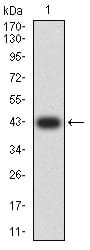
Figure 2:Western blot analysis using BCL11B mAb against human BCL11B (AA: 1-150) recombinant protein. (Expected MW is 42.3 kDa)
Western Blot
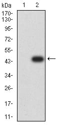
Figure 3:Western blot analysis using BCL11B mAb against HEK293 (1) and BCL11B (AA: 1-150)-hIgGFc transfected HEK293 (2) cell lysate.
Immunofluorescence analysis
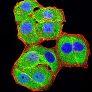
Figure 4:Immunofluorescence analysis of Hela cells using BCL11B mouse mAb (green). Blue: DRAQ5 fluorescent DNA dye. Red: Actin filaments have been labeled with Alexa Fluor- 555 phalloidin. Secondary antibody from Fisher (Cat#: 35503)
Flow cytometric
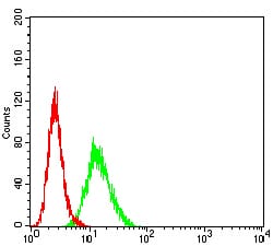
Figure 5:Flow cytometric analysis of Hela cells using BCL11B mouse mAb (green) and negative control (red).
Flow cytometric
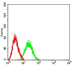
Figure 6:Flow cytometric analysis of Jurkat cells using BCL11B mouse mAb (green) and negative control (red).
Immunohistochemical analysis
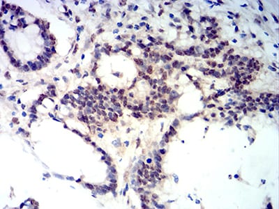
Figure 7:Immunohistochemical analysis of paraffin-embedded colon cancer tissues using BCL11B mouse mAb with DAB staining.
Immunohistochemical analysis
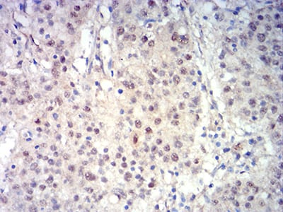
Figure 8:Immunohistochemical analysis of paraffin-embedded stomach cancer tissues using BCL11B mouse mAb with DAB staining.
For Research Use Only. Not for use in diagnostic procedures.