
BACE1 Primary Antibody
Item Information
Catalog #
Size
Price
Description
Cerebral deposition of amyloid beta peptide is an early and critical feature of Alzheimer's disease. Amyloid beta peptide is generated by proteolytic cleavage of amyloid precursor protein (APP) by two proteases, one of which is the protein encoded by this gene. The encoded protein, a member of the peptidase A1 protein family, is a type I integral membrane glycoprotein and aspartic protease that is found mainly in the Golgi. Multiple transcript variants encoding different isoforms have been described for this gene.
Product Overview
Entrez GenelD
23621
Aliases
ASP2; BACE; HSPC104
Clone#
3C1C3
Host / Isotype
Mouse / IgG1
Species Reactivity
Human, Mouse, Rat
Immunogen
Purified recombinant fragment of human BACE1 (AA: 112-324) expressed in E. Coli.
Formulation
Purified antibody from tissue culture in PBS with 0.05% sodium azide
Storage
Store at 4°C short term. Aliquot and store at -20°C long term. Avoid freeze/thaw cycles.
Product Applications
WB (Western Blot)
1/500 - 1/2000
ICC (Immunocytochemistry)
1/200 - 1/1000
FCM (Flow Cytometry)
1/200 - 1/400
ELISA
1/10000
References
1. J Neurochem. 2012 Jan;120 Suppl 1:62-70.
2. Eur J Neurosci. 2010 Oct;32(7):1223-38.
2. Eur J Neurosci. 2010 Oct;32(7):1223-38.
Product Image
Western Blot
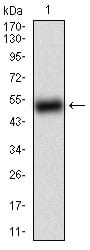
Figure 1: Western blot analysis using BACE1 mAb against human BACE1 (AA: 112-324) recombinant protein. (Expected MW is 49.9 kDa)
Western Blot

Figure 2: Western blot analysis using BACE1 mAb against HEK293 (1) and BACE1 (AA: 112-324)-hIgGFc transfected HEK293 (2) cell lysate.
Western Blot
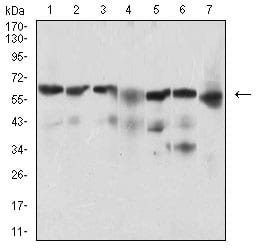
Figure 3: Western blot analysis using BACE1 mouse mAb against Hela (1), SK-N-SH (2), HepG2 (3), C6 (4), PC-12 (5), PANC-1 (6), NIH/3T3 (7) cell lysate.
Immunofluorescence analysis

Figure 4: Immunofluorescence analysis of MCF-7 cells using BACE1 mouse mAb (green). Blue: DRAQ5 fluorescent DNA dye. Red: Actin filaments have been labeled with Alexa Fluor-555 phalloidin. Secondary antibody from Fisher (Cat#: 35503)
Flow cytometric
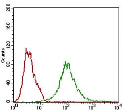
Figure 5: Flow cytometric analysis of Hela cells using BACE1 mouse mAb (green) and negative control (red).
Elisa
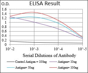
Black line: Control Antigen (100 ng); Purple line: Antigen(10ng); Blue line: Antigen (50 ng); Red line: Antigen (100 ng);
For Research Use Only. Not for use in diagnostic procedures.