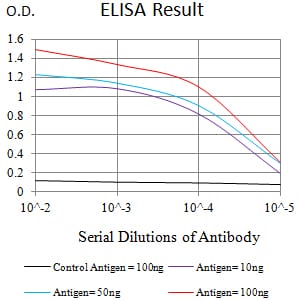
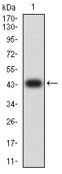
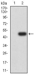
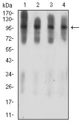
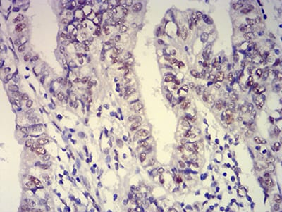
ATXN1 Primary Antibody

Item Information
Catalog #
Size
Price
Description

The autosomal dominant cerebellar ataxias (ADCA) are a heterogeneous group of neurodegenerative disorders characterized by progressive degeneration of the cerebellum, brain stem and spinal cord. Clinically, ADCA has been divided into three groups: ADCA types I-III. ADCAI is genetically heterogeneous, with five genetic loci, designated spinocerebellar ataxia (SCA) 1, 2, 3, 4 and 6, being assigned to five different chromosomes. ADCAII, which always presents with retinal degeneration (SCA7), and ADCAIII often referred to as the `pure' cerebellar syndrome (SCA5), are most likely homogeneous disorders. Several SCA genes have been cloned and shown to contain CAG repeats in their coding regions. ADCA is caused by the expansion of the CAG repeats, producing an elongated polyglutamine tract in the corresponding protein. The expanded repeats are variable in size and unstable, usually increasing in size when transmitted to successive generations. The function of the ataxins is not known. This locus has been mapped to chromosome 6, and it has been determined that the diseased allele contains 40-83 CAG repeats, compared to 6-39 in the normal allele, and is associated with spinocerebellar ataxia type 1 (SCA1). Alternative splicing results in multiple transcript variants, with one variant encoding multiple distinct proteins, ATXN1 and Alt-ATXN1, due to the use of overlapping alternate reading frames.

Product Overview
Entrez GenelD
6310
Aliases
ATX1; SCA1; D6S504E
Clone#
4C7B11
Host / Isotype
Mouse / IgG1
Species Reactivity
Human, Mouse, Rat, Monkey
Immunogen
Purified recombinant fragment of human ATXN1 (AA: 645-815) expressed in E. Coli.
Formulation
Purified antibody in PBS with 0.05% sodium azide
Storage
4°C; -20°C for long term storage
Product Applications
WB (Western Blot)
1/500 - 1/2000
IHC_P(Immunohistochemistry)
1/200 - 1/1000
FCM (Flow Cytometry)
1/200 - 1/400
ELISA
1/10000
References
1.Oncotarget. 2017 Mar 14;8(11):18248-18259.
2.One. 2013 Oct 14;8(10):e76456.
Product Image
Elisa
Figure 1:Black line: Control Antigen (100 ng);Purple line: Antigen (10ng); Blue line: Antigen (50 ng); Red line:Antigen (100 ng)
Western Blot
Figure 2:Western blot analysis using ATXN1 mAb against human ATXN1 (AA: 645-815) recombinant protein. (Expected MW is 44.1 kDa)
Western Blot
Figure 3:Western blot analysis using ATXN1 mAb against HEK293 (1) and ATXN1 (AA: 645-815)-hIgGFc transfected HEK293 (2) cell lysate.
Western Blot
Figure 4:Western blot analysis using ATXN1 mouse mAb against C6 (1), COS7 (2), NIH/3T3 (3), and HL-60 (4) cell lysate.
Flow cytometric
Figure 5:Flow cytometric analysis of Jurkat cells using ATXN1 mouse mAb (green) and negative control (red).
Immunohistochemical analysis
Figure 6:Immunohistochemical analysis of paraffin-embedded endometrial cancer tissues using ATXN1 mouse mAb with DAB staining.
For Research Use Only. Not for use in diagnostic procedures.