
ATPIF1 Primary Antibody
Item Information
Catalog #
Size
Price
Description
This gene encodes a mitochondrial ATPase inhibitor. Alternative splicing occurs at this locus and three transcript variants encoding distinct isoforms have been identified.
Product Overview
Entrez GenelD
93974
Aliases
IP; ATPI; ATPIP
Clone#
1F3B8
Host / Isotype
Mouse / IgG2b
Species Reactivity
Human
Immunogen
Purified recombinant fragment of human ATPIF1 (AA: 1-106) expressed in E. Coli.
Formulation
Purified antibody in PBS with 0.05% sodium azide
Storage
Store at 4°C short term. Aliquot and store at -20°C long term. Avoid freeze/thaw cycles.
Product Applications
WB (Western Blot)
1/500 - 1/2000
IHC_P(Immunohistochemistry)
1/200 - 1/1000
ICC (Immunocytochemistry)
1/100 - 1/500
FCM (Flow Cytometry)
1/200 - 1/400
ELISA
1/10000
References
1.Pathobiology. 2015;82(5):224-32.
2.Cell Rep. 2014 Apr 10;7(1):27-34.
2.Cell Rep. 2014 Apr 10;7(1):27-34.
Product Image
Elisa

Figure 1: Black line: Control Antigen (100 ng);Purple line: Antigen (10ng); Blue line: Antigen (50 ng); Red line:Antigen (100 ng)
Western Blot

Figure 2:Western blot analysis using ATPIF1 mAb against human ATPIF1 (AA: 1-106) recombinant protein. (Expected MW is 38.2 kDa)
Western Blot

Figure 3:Western blot analysis using ATPIF1 mAb against HEK293 (1) and ATPIF1 (AA: 1-106)-hIgGFc transfected HEK293 (2) cell lysate.
Immunofluorescence analysis

Figure 4:Immunofluorescence analysis of Hela cells using ATPIF1 mouse mAb (green). Blue: DRAQ5 fluorescent DNA dye. Red: Actin filaments have been labeled with Alexa Fluor- 555 phalloidin. Secondary antibody from Fisher (Cat#: 35503)
Flow cytometric
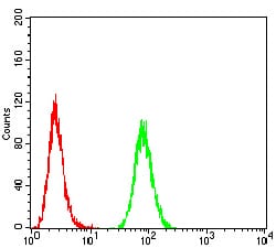
Figure 5:Flow cytometric analysis of Hela cells using ATPIF1 mouse mAb (green) and negative control (red).
Immunohistochemical analysis

Figure 6:Immunohistochemical analysis of paraffin-embedded lung cancer tissues using ATPIF1 mouse mAb with DAB staining.
Immunohistochemical analysis

Figure 7:Immunohistochemical analysis of paraffin-embedded cervical cancer tissues using ATPIF1 mouse mAb with DAB staining.
For Research Use Only. Not for use in diagnostic procedures.